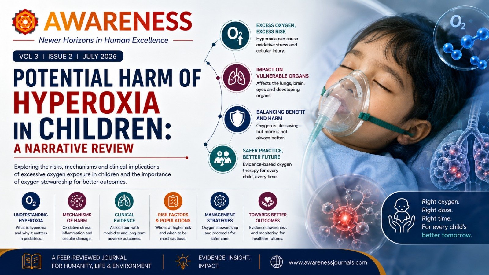
Potential Harm of Hyperoxia in Children: A Narrative Review
- Aug 10, 2026
- 260 views
Abstract
Background: Hyperoxia is common in pediatric critical care, yet its harmful effects remain underappreciated. This review combines clinical, physiologic, and molecular evidence showing that excessive oxygen exposure causes a range of lung and systemic injuries.
Methods: A structured literature search was performed in PubMed, Scopus, and Embase. Articles were selected to represent mechanistic, translational, and clinical evidence across relevant pediatric populations. The initial search strategy yielded 252 articles; 194 studies were excluded after initial screening. The remaining studies underwent independent full-text review and data extraction.
Pathophysiology: High FiO2 leads to oxidative stress, damage to alveolar epithelial and endothelial cells, increased reactive oxygen species, release of inflammatory cytokines, changes in gene expression, and disruption of the alveolar-capillary barrier. Hyperoxia contributes to atelectasis, reduced lung compliance, decreases in vital capacity and diffusing capacity, and, with prolonged exposure, diffuse pneumonitis. Beyond the lungs, hyperoxia impairs immune regulation, gastrointestinal integrity, and nervous system development. Results: Clinical research shows mixed results across different populations, but severe or sustained hyperoxemia is consistently linked to higher morbidity and mortality in critically ill children, those on ECMO, and certain neonatal and trauma groups. Studies in pediatric cardiac arrest and traumatic brain injury highlight the complex relationship between oxygen levels and neurologic outcomes, with hypoxia showing the strongest link to death. Advances like automated, closed-loop FiO2 controllers show promise for optimizing oxygen delivery and reducing exposure to harmful extremes. Emerging molecular therapies, including growth factor modulation, antioxidants, and stem-cell-based treatments, may further reduce oxygenrelated injury.
Conclusion: Overall, the evidence underscores the importance of carefully managing oxygen, a powerful drug with both lifesaving benefits and significant risks. Keywords: Hyperoxia, hypoxia, critical care, pediatrics, oxygen toxicity Corresponding Author: Vidit Bhargava, vbhargava@uabmc.edu
Full Text
1. Introduction
Oxygen is a common therapy to treat hypoxia in patients. Although vital to cell function, excessive exposure to oxygen can be detrimental.1 While hyperbaric oxygen has specific clinical indications, excessive oxygen administration in most other conditions is unwarranted and often underrecognized.2 While exposure to high levels of oxygen has been well-studied in animals, the clinical relevance of these findings may not be fully applicable to humans, as different species respond differently to high oxygen exposure.3 In this review, we discuss results from human clinical and in vitro studies regarding the pulmonary effects of excessive oxygen administration.
2. Materials and Methods
A structured literature search was performed in PubMed, Scopus, and Embase using combinations of the terms: hyperoxia, oxygen toxicity, pediatric, PICU, ECMO, traumatic brain injury, cardiac arrest, perioperative oxygen, COVID-19, automated FiO2 control. Articles were selected to represent mechanistic, translational, and clinical evidence across relevant pediatric populations. Because of marked heterogeneity in patient groups, exposures, and outcomes, formal systematic review methodology (PRISMA/PROSPERO) was not appropriate, and no meta-analysis was attempted. The initial search strategy yielded 252 articles, which were downloaded to Covidence for further screening by two reviewers (VB, JC). 194 studies were found irrelevant based on the inclusion and exclusion criteria. The remaining studies underwent independent full-text review and data extraction by the two reviewers (VB, JC) based on the inclusion and exclusion criteria listed here.
Inclusion Criteria: Case series, case control, cohort, clinical trial (randomized or non-randomized), meta-analysis with or without review. Population: pediatric, neonate, lab studies focused on hyperoxia. Intervention/exposure: exposure to oxygen via mechanical or non-mechanical routes, lab model of hyperoxia. Outcomes: measures of cell, tissue, or organ dysfunction.
Exclusion Criteria: Case report, review without meta-analysis, protocols. Population: adults. Intervention/exposure: if no exposure to oxygen. Outcomes: if no outcome measures are studied.
3. Definitions
The term hyperoxia is used interchangeably to describe both excessive oxygen administration as well as elevated partial pressure of oxygen (PaO2) in the body. Excessive oxygen delivery and hyperoxemia can occur independently. In the setting of severe lung disease and limited diffusion, the PaO2 may remain low or normal. Therefore, while pulmonary oxygen toxicity may occur with high oxygen delivery, hyperoxemia may not. On the other hand, any FiO2 >21% has the potential to cause hyperoxemia and toxicity in organs exposed to elevated PaO2.
Better terminology is necessary to characterize the appropriateness of oxygen therapy. Excessive oxygen delivery is the amount of administered oxygen that causes oxygen toxicity. Hyperoxemia refers to the presence of a supraphysiologic amount of oxygen in the blood, commonly defined by a PaO2 threshold. The specific threshold of the fraction of inspired oxygen (FiO2) above which toxicity occurs depends on the cell tolerance of the concentration and duration of oxygen to which it is exposed. Different cell types have different thresholds and variable responses.4 Pulmonary oxygen toxicity is most common during normobaric oxygen therapy as airway and alveolar epithelial cells have the most exposure. A definitive threshold concentration or duration of oxygen therapy has not been found in humans. The commonly cited FiO2 limit of 50–60% is largely based on animal and in vitro studies.5,6 A clear threshold PaO2 that leads to end-organ injury has also not been identified. Many studies use PaO2 thresholds beyond (e.g., 300 mm Hg) the upper limit of normal (approximately 100 mm Hg). Further studies are necessary to define the limits of oxygen therapy and toxicity in humans. Until more is known, setting FiO2 with target oxygen saturation and PaO2 ranges (with upper limits) is most appropriate, with the acknowledgment that pulmonary toxicity can occur at high FiO2 settings. Recently published pediatric guidelines in the management of patients with acute respiratory distress syndrome (ARDS) recommend maintaining a goal SpO2 88%–97% and <92% in patients with severe ARDS with central venous oxygen saturation monitoring.7
4. Pathophysiology
Pathological changes occur with excessive oxygen delivery. In the 70s, multiple post-mortem case series attributed hyaline membrane formation and capillary proliferation in the child and adult population to high oxygen concentrations. Across both pediatric and adult patients—many without preexisting lung disease—post-mortem and clinical observations revealed diffuse alveolar damage after sustained oxygen therapy, regardless of delivery method or duration. Many patients in studies showing hyaline membrane disease had primary or secondary lung disease, required mechanical ventilation with high or unknown volumes or pressures, and received variable durations of oxygen supplementation. Collectively, these reports established an association between excessive FiO2 and structural lung injury, though confounding factors such as infection and ventilation parameters make it difficult to attribute causality solely to oxygen toxicity.
In mechanically ventilated patients with acute lung injury (ALI), excessive oxygen delivery—e.g., FiO2 >50% with SpO2 >92%—is common and linked to adverse changes.8 For example, FiO2 1.0 increases intrapulmonary shunting due to derecruitment resulting from resorption atelectasis.9 Pathological changes due to hyperoxia in mechanically ventilated patients are similar to those in patients who died with acute respiratory distress syndrome. Diffuse alveolar damage can occur after 48 hours of exposure to elevated oxygen levels.10 During the exudative phase of ARDS, alveolar epithelial and endothelial cells show increased ROS-generating enzymes and apoptosis signaling (including NOX-linked pathways), consistent with concurrent oxidative stress and programmed cell death.11 Even though oxygen toxicity cannot be identified as a cause of ALI, it can certainly contribute to similar clinical findings and exacerbate the disease process.
While many of the negative effects of excessive oxygen exposure on the lungs are well-documented, it is still unclear just how long it takes before this exposure causes significant risks. According to published reports, 6 hours of elevated oxygen increases reactive oxygen species and the elevation of inflammatory markers, a decrease in vital capacity occurs between ~24–77 hours on FiO2 0.75–1.0, and tracheobronchitis occurs after ~6 hours on FiO2 1.0 or ~12 hours on FiO2 0.75.12,13 Although the duration of hyperoxia in these studies is arbitrary, we can estimate that injury begins within hours of exposure and progresses over days, with a likely cumulative effect on lung architecture and mechanics.
Molecular Pathophysiology
Oxygen toxicity results from interference of reactive oxygen species (ROS) with normal cell structure and function and can occur with excessive oxygen exposure. ROS include superoxide (O2−·), hydrogen peroxide (H2O2), and hydroxyl (·OH). They can form without excessive oxygen delivery and under physiologic conditions (e.g., infection). Normally, enzymes such as superoxide dismutase and glutathione peroxidase inactivate reactive oxygen species. However, when antioxidant mechanisms are overwhelmed, such as in critical illness, oxygen toxicity can occur. ROS are produced by various cells through enzymes such as NADPH oxidase and xanthine oxidase.11 Phagocytic cells (e.g., neutrophils, macrophages), pulmonary vascular endothelial cells, and alveolar epithelial cells have all been implicated in the production of ROS.11,14 NADPH oxidase is an important source of ROS in pulmonary endothelial cells and alveolar epithelial cells.11,14 The SphK1/S1P/Spns2 axis also drives hyperoxia-induced endothelial NOX2 activation and ROS generation.15
ROS cause cellular injury by altering gene expression, signaling pathways, and/or damaging DNA, protein, and lipids.16 Cell signaling is altered to initiate protective mechanisms, such as maintaining mitochondrial integrity or triggering programmed cell death, during oxidative stress.17 Increase in ATP production in human pulmonary endothelial cells and release into the extracellular environment leads to activation of receptors that signal changes in protein synthesis and glucose metabolism during periods of oxidative stress.18 Enzymes involved with apoptosis increased in airway epithelial cells exposed to >95% oxygen in vitro for up to 72 hours. These enzymes induced cell death by affecting apoptotic signaling pathways.19 Alveolar epithelial function is also affected by changes to signaling pathways that lead to cell death, evidenced by apoptotic and necrotic changes. Other signaling mechanisms can enhance the ROS response, possibly activating protective mechanisms. Activation of mitogen-activated protein kinases (MAPK) in human pulmonary endothelial cells occurred when exposed to 95% oxygen.14 MAPKs, which are important signaling proteins, in turn alter signaling pathways that increased ROS production.
Altered gene expression in the lungs results in the synthesis of proteins that regulate detoxification, inflammation, cell proliferation, and cell death. Although these changes are initially adaptive—aimed at protecting cells from oxidative injury—they can paradoxically contribute to local damage through inflammation and cell loss, ultimately impairing lung function. In vitro studies have shown that exposing human pulmonary artery endothelial cells to 90% oxygen for 48–72 hours increases the expression of intercellular adhesion molecule-1 (ICAM-1), thereby enhancing neutrophil adherence.20 Similarly, exposure to hyperoxia modulates the expression of pro-inflammatory cytokines, including TNF-α, IL-1β, IL-6, and IL-8, with IL-8 demonstrating the most consistent and pronounced increase. In alveolar macrophages isolated from bronchoalveolar lavage (BAL) fluid of children and exposed to 95% oxygen for 48 hours, IL-8 levels were significantly elevated. A comparable increase in IL-8 was also associated with enhanced polymorphonuclear leukocyte chemotaxis in adult cell models. Inflammatory activation also extends to TNF-α; its concentration rises under both LPS-stimulated and high-oxygen conditions, reaching statistically significant levels in macrophages exposed to 60–95% oxygen compared with normoxic (21%) conditions.21 Additional genes implicated in oxygen-induced injury include gasdermin D (GSDMD), which mediates pyroptotic cell death, and connective tissue growth factor (CTGF), which promotes fibrotic and inflammatory pathways.22,23
Excessive oxygen leads to short and long-term damage. Alveolar macrophages release fibronectin and alveolar-macrophage-derived growth factors, which attract fibroblasts, causing chronic lung changes. Human lung fibroblasts exposed to 95% oxygen ROS increased RhoA activation.24 RhoA is a small GTPase that regulates various cell functions, including the mediation of collagen-I synthesis and lung fibrosis in the setting of high oxygen.
While most studies investigating the molecular pathophysiology of hyperoxia have focused on cell signaling in the lungs (and, in the neonatal population, retinal tissues), there is also evidence of damage to various other organs including the brain, immune system, and gastrointestinal tract. Yis et al found that exposure of rat pups to 80% oxygen from birth to five days of life was associated with increased cell death in the developing brain as compared to 21% oxygen.25 Additional studies have shown impaired FoxP3+-regulated T cell generation and T cell autoreactivity in mice, increased secretory IgA (SIgA) from intestinal epithelial cells, and gut inflammation/dysbiosis via TLR-4, TNF, NF-κB, and Nrf2 pathway activation.22,25,26
Alveolar Cell Death & Damage to Alveolar-Capillary Barrier
ROS-induced changes in gene expression, signaling pathways, and damage to DNA, proteins, and lipid membranes can lead to cell death by apoptosis and/or necrosis. Many studies support apoptotic processes in cells exposed to high levels of oxygen. Apoptosis was attenuated in the presence of vitamin E, an antioxidant. Evidence of necrosis was observed in alveolar epithelial cells, specifically in human lung adenocarcinoma cells A549, upon exposure to 95% oxygen. The term “necroptosis” has been proposed to encompass a dual mechanism of cell death. Simultaneous occurrence in different cells and/or sequential processes has been suggested. Injury to alveolar epithelial and endothelial cells compromises the alveolar-capillary barrier. This occurs independent of cellular inflammation when exposed to excessive oxygen.
5. Clinical Studies
Early studies dating back to the 1940s showed the effects of the delivery of high concentrations of oxygen to the lungs. A few studies isolated excessive oxygen exposure by including only healthy adults with no history of lung pathology and administering oxygen by noninvasive methods, thereby excluding any effects of positive-pressure ventilation. Later studies included mechanically ventilated lungs. Many of these studies did not provide details on ventilator management or explain the possible roles of barotrauma or volutrauma. Accurate conclusions cannot be made about the isolated effects of oxygen therapy, but the results indicate that caution is warranted when determining the level of FiO2 to administer.
Hyperoxia Clinical Trials
Elevated oxygen levels, when employed clinically, can induce various pulmonary damages, ranging from tracheobronchitis to widespread alveolar injury. Tracheobronchitis, often attributable to diminished ciliary activity, represents among the earliest indicators of pulmonary oxygen toxicity. Patients may present with chest pain, cough, and dyspnea.13 For instance, in one study, the exposure to 95% oxygen via a non-rebreather mask for 15.5–18 hours resulted in chest pain in 9 of 14 patients.27 Although the discomfort subsided following cessation of oxygen therapy, bronchoscopy revealed erythema in 6 patients, indicative of inflammation.
Further symptoms of pulmonary oxygen toxicity include alveolar atelectasis (both absorptive and obstructive), decreased lung compliance, reduced vital capacity, and diminished diffusing capacity.28 A study involving eight aircrew subjected to high oxygen delivery and hypergravity associated oxygen exposure with atelectasis, with ultrasound indicating greater atelectasis following breathing 100% oxygen compared to 44.5% and 21% oxygen.29 A decline in vital capacity was noted between 24 and 77 hours at FiO2 0.75–1.0. Although limited, these studies suggest that oxygen levels of 50% or higher can provoke adverse clinical effects, with cellular damage likely initiating within hours, even if clinical signs manifest subsequently.
Extensive research has examined hyperoxia’s impact on mortality across various populations. These are summarized below in Table 1.
Table 1: Characteristics and Outcomes of Studies Assessing Hyperoxia in Critically Ill ICU Populations
| Author, Year | Target Population | Study Design, N | Oxygenation Parameter | Study Findings |
|---|---|---|---|---|
| Kapadia, 201340 | Neonates (24–34 weeks GA); delivery room resuscitation | Prospective RCT; N = 88 (44 per group) | Limited oxygen strategy (LOX): incremental increase in FiO2 from 21% to target SpO2. High oxygen strategy (HOX): 100% FiO2 initiated and adjusted every 30 s to meet target SpO2 85–94%. | LOX effectively resuscitated preterm neonates, decreased oxygen load by 50% (401 ± 151 vs 662 ± 249; P < .01), lowered ventilator days (3 [0–64] vs 8 [0–96]; P < .05), rescue HFOV, and BPD (7% vs 25%; P < .05). LOX group spent less time with SpO2 > 94% and developed less oxidative stress by 1 hour of life. |
| Lilien, 202241 | Pediatric patient | Systematic review N = 27,555; 11 studies pooled for meta-analysis (N = 23,204) | Variable definitions of hyperoxia, from PaO2 > 120 mm Hg to > 300 mm Hg. | Variability in mortality associated with hyperoxia across studies. Hyperoxia increased the odds of mortality (OR 1.59, 95% CI 1.00–2.51). |
| Ramgopal, 201932 | Pediatric ICU patients (< 30 days to ≤ 18 years) with PaO2 at any point during admission | Retrospective cohort study; N = 6,250 | Severe hyperoxemia: PaO2 ≥ 300 mm Hg. | Severe hyperoxemia (dose-response) independently associated with in-hospital mortality (aOR 1.78; 95% CI 1.36–2.33; P < .001). |
| Ramgopal, 202033 | PICU patients with PaO2 obtained 6 h prior to or after admission | Single-center observational study; N = 4,093 | Hyperoxemia: PaO2 ≥ 300 mm Hg; Hypoxia: PaO2 < 60 mm Hg. | Hypoxemia associated with increased in-hospital mortality. Hyperoxemia overall not associated with mortality (aOR 1.38; 95% CI 0.98–1.93), except at extremes (PaO2 ≥ 550 torr). |
| Raman, 201634 | Patients < 18 years admitted to ICU | Retrospective cohort study; N = 7,410 | Hyperoxia: PaO2 > 300 torr; Hypoxia: PaO2 < 60 torr OR SpO2 < 90%. | U-shaped association between admission PaO2 and mortality. Hypoxia increased risk of death (OR 3.13; 95% CI 1.79–5.48; p < 0.001). No increase with hyperoxia (OR 1.15; 95% CI 0.42–3.17; p = 0.77). |
| Numa, 201837 | Pediatric patients 0–18 years admitted to ICU | Single-center retrospective analysis; N = 1,447 | Values in 50 mm Hg increments. Hyperoxia: PaO2 > 250 mm Hg. | U-shaped relationship between mortality and admission PaO2. Lowest mortality at PaO2 101–150 and 151–200 mm Hg. Highest at PaO2 > 350 and < 50 mm Hg. OR for hyperoxia predicting death 2.66 (p = 0.047). |
| Van der Wal, 202342 | Patients > 18 years, mechanical ventilation ≥ 24 h | Randomized multicenter trial; N = 664 (stopped early due to COVID-19; 664 of planned 1,512) | Low: PaO2 55–80 mm Hg OR SpO2 91–94%. High: PaO2 110–150 mm Hg OR SpO2 96–100%. | No reduction in 28-day mortality with low oxygenation vs high oxygenation strategy. |
| Balcarcel, 202231 | Patients < 18 years requiring mechanical ventilation within 3 days of admission | Retrospective observational cohort study; N = 5,406 | Hypoxemia: SpO2 < 90% in first 24 h. CEOE: mean hourly FiO2 above 0.21 when SpO2 ≥ 95% during first 24 h. | Higher CEOE increased odds of in-hospital mortality (aOR 1.7; 95% CI 1.1–2.9) and MODS on day 7 (aOR 3.9; 95% CI 2.7–5.9). Hypoxemia-associated mortality and MODS comparable to highest CEOE group. |
| Geva, 202336 | Patients 7 months to < 16 years, PICU, ventilated via ETT ≥ 24 h | Retrospective cross-sectional study; N = 3,354 | Hypoxia: SpO2 < 90% in 24 h. Hyperoxia: CEOE mean hourly FiO2 above 0.21 when SpO2 ≥ 95% during first 24 h. | Higher CEOE quartiles associated with greater mortality, primarily in the highest quartile. |
Abbreviations: randomized control trial (RCT), fraction of inspired oxygen (FiO2), arterial partial pressure of oxygen (PaO2), oxygen peripheral saturation (SpO2), odds ratio (OR), intensive care unit (ICU), adjusted odds ratio (aOR), pediatric intensive care unit (PICU), high frequency oscillatory ventilation (HFOV), bronchopulmonary dysplasia (BPD), cumulative excess oxygen exposure (CEOE), multiorgan dysfunction syndrome (MODS).
Hyperoxia and Critically Ill Pediatric Patients
Hyperoxia is common in critically ill children at admission to a PICU and is associated with increased risk-adjusted mortality. The effect of hyperoxia on critically ill pediatric patients appears dose-dependent. Like the studies on cardiac arrest, it is consistently observed that hypoxia results in worse outcomes compared to hyperoxia. However, several studies have identified a link between hyperoxia and increased morbidity and mortality in critically ill children. Most pediatric research indicates that severe hyperoxia or hyperoxemia is associated with poorer outcomes, especially with longer or more frequent exposures to these conditions.30–36 The level of hyperoxia differs across studies, with Numa et al identifying a risk minimum around a PaO2 of 100–200 mmHg, whereas Pelletier et al reported that harmful hyperoxia occurs over 450 mmHg in one study and between 250–400 mmHg in another.37–39 (Table 1)
Hyperoxia and Traumatic Brain Injury (TBI)
Numerous studies involving both adult and pediatric TBI patients suggest that hyperoxia might be neuroprotective. While some studies suggest improvement in biomarkers and reduced intracranial pressure across adult and pediatric patients, none of the studies demonstrated an association between hyperoxia and improvement in neurological outcomes. (Table 2)
Table 2: Characteristics and Outcomes of Studies Assessing Hyperoxia in Critically Ill Patients After Trauma or Traumatic Brain Injury
| Author, Year | Target Population | Study Design, N | Oxygenation Parameter | Study Findings |
|---|---|---|---|---|
| Baekgaard, 202043 | Trauma patients > 17 years | Observational study; multicenter prospective trauma registry; N = 5,912 | Hyperoxia: PaO2 ≥ 150 mmHg on admission. | Univariate analysis showed higher in-hospital mortality for hyperoxemic vs normoxemic patients. After propensity matching, significantly lower in-hospital mortality for hyperoxemic patients. |
| Jeong, 201844 | Patients > 16 years surviving ≥ 5 days after ED arrival | Observational cohort study; N = 10,141 (exclusions: < 2 ABGs, max PaO2 < 60 mm Hg, death/transfer/complications within 5 days, LOS > 90 days, unable to calculate AUC, acute MI) | AUC over 72 h (AUC72) using highest, average, median PaO2. Hyperoxemia: max > 137, average > 105, median > 103 mm Hg, AUC72 > 174 mm Hg. | Significant correlations between all hyperoxemia variables and 90-day in-hospital mortality. After adjustment, only AUC72 significantly associated (aOR 1.53; 95% CI 1.25–1.88; p < 0.0001). AUC72 also associated with ICU transfer and end-organ failure. |
| Tolias, 200445 | Patients > 16 years, severe TBI, normobaric hyperoxia (FiO2 1.0) for 24 h within 6 h of admission | Prospective clinical trial; N = 52 treatment and N = 112 matched control cohort | Hyperoxia: FiO2 1.0 for 24 h, started within 6 h of admission. | Hyperoxia group: improved biomarker profile and lower intracranial pressure. |
| Nortje, 200846 | Patients > 15 years with TBI | Prospective clinical trial; N = 11 patients | Cerebral microdialysis, brain tissue oximetry (PbO2), and oxygen-15 PET (15O-PET) at normoxia (FiO2 0.35–0.5) and hyperoxia (FiO2 0.6–0.8). Baseline PaO2 99 ± 23; hyperoxia PaO2 226 ± 68. | Hyperoxia increased the mean PbO2 and reduced the lactate:pyruvate ratio. |
| Figaji, 201047 | Patients < 15 years with severe TBI | Prospective observational study; 48 tests in 28 patients with PbO2 monitoring and an oxygen challenge test (temporary FiO2 increase for 15 min) | Pre- and post-test values (PbO2, ICP, CPP, PaO2, SaO2, PaCO2) measured. | Linear increase in PbO2 with normobaric hyperoxia. A greater PbO2 response to PaO2 change is associated with a worse outcome. |
| Ketharanathan, 202048 | Patients < 18 years with accidental severe TBI (GCS ≤ 8) | Retrospective explorative study; N = 71 | Hyperoxia cutoffs: PaO2 > 200, > 250, > 300 mm Hg. Highest PaO2 in first 24 h; cumulative AUC cutoffs: < 2000 (physiological), 2000–4000 (intermediate), > 4000 (high). | Hyperoxia classification differed by cutoff vs AUC analysis. AUC better approximates physiological circumstances via a time- and dose-dependent approach. |
| Tang, 202449 | Patients < 18 years with severe accidental TBI (GCS 3–8) | Prospective institutional registry; N = 98 | First, highest, and AUC PaO2 in first 24 h evaluated. Hyperoxia: PaO2 > 300 mm Hg. | Hyperoxia incidence 33%. Hyperoxia not associated with unfavorable outcome after 6 months. |
Abbreviations: arterial partial pressure of oxygen (PaO2), emergency department (ED), arterial blood gas (ABG), length of stay (LOS), area under the curve (AUC), myocardial infarction (MI), area under the curve over 72 hours (AUC72), adjusted odds ratio (aOR), confidence interval (CI), traumatic brain injury (TBI), fraction of inspired oxygen (FiO2), brain tissue oximetry (PbO2), intracranial pressure (ICP), cerebral perfusion pressure (CPP), arterial oxygen saturation (SaO2), arterial partial pressure of carbon dioxide (PaCO2), Glasgow coma score (GCS).
Hyperoxia and Pediatric Cardiac Arrest
Multiple studies have examined the link between hyperoxia and outcomes following pediatric cardiac arrest. These are summarized below in Table 3. The smaller studies demonstrated no difference in outcomes in patients with hyperoxia (PaO2 > 200 mmHg) in the first six hours.50 The larger studies revealed worse outcomes associated with hypoxia and extremes of hyperoxia (PaO2 > 600 mmHg), which the studies concluded were rarely observed.51 These studies concluded that in the post-cardiac-arrest phase hypoxia is more critical than hyperoxia and associated with worse outcomes.
Table 3: Characteristics and Outcomes of Studies Assessing Hyperoxia in Critically Ill Patients After Cardiac Arrest or Requiring Extracorporeal Membrane Oxygenation (ECMO)
| Author, Year | Target Population | Study Design, N | Oxygenation Parameter | Study Findings |
|---|---|---|---|---|
| Sznycer-Taub, 201652 | Patients < 1 year undergoing cardiac surgery requiring postoperative VA ECMO | Retrospective chart review; N = 93 | Hyperoxia: mean PaO2 > 193 mm Hg in the first 48 h of ECMO. | Total mortality ~38%. Hyperoxia an independent risk factor for 30-day mortality (p = 0.001) and associated with the need for dialysis (p = 0.02). |
| Bennett, 201350 | Pediatric patients 24 hours to 18 years with cardiac arrest and return of circulation ≥ 20 min | Retrospective cohort study; N = 195 cardiac arrest events | Hyperoxia: PaO2 > 200 mm Hg in the first 6 h post-arrest. Hypoxia: PaO2 < 50 mm Hg. | 13% maintained both normoxia and normoventilation; 10% had both hyperoxia and hypoxia. No difference in outcomes (survival with good outcome 34% for hyperoxia vs 42% normoxia; p = 0.23). |
| Ferguson, 201251 | Patients < 16 years with cardiac arrest and ABG within 1 h of PICU admission | Retrospective cohort study; N = 1,875 | Hypoxia: PaO2 < 60 mm Hg; Hyperoxia: PaO2 ≥ 300 mm Hg. | Mortality 39%. Incidence: hypoxia 24%, hyperoxia 11%, normoxia 65%. Severe hypoxia increased death probability (OR 1.92; 95% CI 1.80–2.21). Hyperoxia also increased ICU mortality risk (OR 1.2; 95% CI 1.0–1.5; OR 1.12 per 100 mm Hg increase in PaO2). Optimal PaO2: 60–75 mm Hg. |
| Cashen, 201835 | Pediatric patients < 19 years treated with ECMO | Retrospective, secondary analysis; N = 484 | Hyperoxia: highest PaO2 > 200 Torr during the first 48 h on ECMO. | Hyperoxia associated with higher mortality and shorter durations of ECMO and ICU stays (167 [50.5%] vs 48 [31.4%]; p < 0.001). No functional status differences at discharge. |
| Frazier, 202453 | Patients ≤ 18 years receiving chest compressions during ICU stay | Embedded prospective observational study; N = 284. Exposures based on lowest PaO2 within first 24 h post-arrest | Hypoxemia: PaO2 < 60 mm Hg; Hyperoxemia: PaO2 ≥ 300 mm Hg; Normoxemia: PaO2 60–300 mm Hg. | Hypoxemia group less likely to survive to hospital discharge (aRR 0.71; 95% CI 0.58–0.87). |
Abbreviations: veno-arterial extracorporeal membrane oxygenation (VA ECMO), arterial partial pressure of oxygen (PaO2), arterial blood gas (ABG), pediatric intensive care unit (PICU), intensive care unit (ICU), odds ratio (OR), adjusted risk ratio (aRR).
6. Hyperoxia in COVID-19
Hyperoxemia was common during the COVID-19 pandemic, especially among mechanically ventilated patients receiving prolonged high FiO2.54 Prospective and observational cohorts consistently showed a high prevalence of hyperoxemia and “excessive oxygen use” in COVID-19 ARDS populations. Outcome associations, however, have been inconsistent. In a single-center cohort of invasively ventilated patients, time spent in hyperoxemia (PaO2 > 100 mmHg) and combined hyperoxia–hyperoxemia (FiO2 > 0.60 with PaO2 > 100 mmHg) were independently linked to higher ICU mortality and increased ventilator-associated pneumonia.55 Conversely, analysis of the multicenter PRoVENT-COVID study found no difference in 28-day, 90-day, or out-of-hospital mortality between hyperoxemic and normoxemic patients after matching, despite frequent early PaO2 > 90 mmHg.54 Interventional evidence from the HOT-COVID randomized trial showed that targeting a lower PaO2 (≈60 mmHg) was associated with more days alive without life support than targeting a higher PaO2 (≈90 mmHg), supporting conservative oxygen strategies, even without a difference in mortality.56 Overall, these data suggest that while hypoxemia remains the primary concern, sustained hyperoxemia offers no benefit and may cause harm, emphasizing the importance of early FiO2 reduction in COVID-19 ARDS. There is currently a lack of specific pediatric data on hyperoxemia (e.g., elevated PaO2 levels) and its effect on clinical outcomes in children with COVID-19. Current pediatric COVID-19 research mainly concentrates on respiratory support methods, predictors of severe disease, and managing hypoxemia, rather than the effects of prolonged high PaO2 exposure.
7. Management Targets
Contemporary pediatric guidance emphasizes that oxygen should be titrated to avoid both hypoxemia and sustained hyperoxemia. The PALICC-2 recommendations for pediatric ARDS support targeting oxygenation ranges that achieve adequate delivery while minimizing FiO2 exposure, using SpO2 goals generally in the 92–97% range and prioritizing early FiO2 reduction once targets are met.7 In pediatric cardiac arrest care, international consensus recommends FiO2 1.0 during active resuscitation, followed by prompt titration after return of spontaneous circulation to avoid extremes of oxygenation (goal SpO2 94–99%); post-ROSC management aims for near-normal oxygenation, avoiding hyperoxemia when arterial blood gases are available.57 Across these settings, guidance consistently highlights frequent reassessment, use of arterial blood gas (ABG) confirmation when feasible, and avoidance of prolonged high FiO2 in the absence of refractory hypoxemia, reflecting the principle that oxygen is a drug with both lifesaving benefits and potential toxicity.
Oxygen administration is similar to other drugs and must be titrated to achieve the desired effect. However, achieving the exact blood oxygen level is challenging. Adjusting FiO2 during maintenance and weaning can be challenging, as even small changes in inspired oxygen can lead to unintended fluctuations in oxygen saturation. Proper titration is essential, but it’s often hindered by factors such as staff experience and staffing levels, leading to significant variability in SpO2 levels. Automated oxygen controllers can help reduce the risks associated with weaning from oxygen therapy. Automated oxygen controllers have successfully maintained oxygen saturation in target ranges in neonatal patients58–65 and shown to be feasible for titration in adults with chronic obstructive pulmonary disease (COPD). Neonatal studies compared manual FiO2 adjustment with automated open and closed-loop FiO2 control systems in neonates receiving both invasive and non-invasive oxygen support. The automated systems consistently demonstrated greater effectiveness in keeping oxygen levels within the target range. While neonatal studies did not consistently prove the prevention of acute hypoxia, they did show a decrease in the duration of prolonged hypoxemic and hyperoxemic episodes.64 Neonatal patients using automated oxygen controllers were found to have much better compliance with normoxic target ranges with less time spent severely hyperoxic.66 Additionally, these patients tended to have a shorter duration of mechanical ventilation compared to those not on automated oxygen controllers.67 These studies are summarized in Table 4.
Table 4: Open- and Closed-Loop Automated Controllers in the Neonatal and Pediatric Population
| Author, Year | Target Population | Study Design, N | Study Findings |
|---|---|---|---|
| IR, 1979 | Preterm neonates in the first week of life | Prospective clinical trial; N = 12. Automated control of FiO2 to meet PaO2 goals 55–80 mm Hg using an indwelling arterial electrode sensor connected to the ventilator. | PaO2 outside target range 12.2% of the time during automatic servo-control vs 27.6% in manual control. |
| Dugdale, 1988 | Neonates with RDS | Prospective clinical trial; N = 7. Microcomputer-based closed-loop control using PaO2 from an indwelling umbilical artery electrode as input; duration 48 h. | Time PaO2 within ± 1 kPa of target using a closed-loop regulator 74.9 ± 10.2% vs 45.2 ± 16.0% with comparable manual control. |
| Bhutani, 1992 | Infants with BPD | Prospective clinical trial; N = 14. Adaptive adjustment of inspired oxygen (FiO2) based on a desired percent arterial hemoglobin saturation. | SpO2 within target range: 54% standard protocol, 69% (P < 0.01) bedside manual control, 81% (P < 0.01) adaptive control. Fewer fluctuations and overshoots with adaptive control. |
| Sun, 1997 | Newborn infants requiring mechanical ventilation | Prospective clinical trial; N = 16. Open-loop computer control of inspired oxygen concentration. | More time at target SaO2 (p < 0.025), SaO2 < 90% less frequent (p < 0.01), and less overall variation than either control period. |
| Claure, 2001 | Mechanically ventilated very low birthweight infants | Prospective clinical trial; N = 14. Closed-loop FiO2 control (cFiO2) vs continuous manual FiO2 (mFiO2) adjustments by a nurse. | cFiO2 noninferior to a fully dedicated nurse in maintaining SpO2 within target range. Mean SpO2 and FiO2 similar. Significant increase in normoxemia duration during cFiO2 (75 ± 13 vs 66 ± 14%). |
| Claure, 2011 | Mechanically ventilated preterm infants | Crossover study; N = 32. Automated vs manual adjustment of FiO2. | Time with SpO2 in goal range increased during automated period (40 ± 14% vs 32 ± 13%), less time hyperoxic. Significant increase in time hypoxic in automated period (32% ± 12% vs 23% ± 9%). |
| Hallenberger, 2014 | Preterm infants on mechanical ventilation or nasal CPAP with supplemental oxygen | Multicenter, randomized controlled, crossover trial; N = 34. Routine manual control of FiO2 vs closed-loop automatic control (CLAC). | Median time with SaO2 within target range 61.4% (31.5–99.5) for routine manual control vs 71.2% (44.0–95.4) for CLAC (p < 0.001). |
| Dijkman, 2025 | Extremely preterm infants surviving to NICU discharge | Prospective cohort study; matched 1:1 with routine manual FiO2 control cohort; N = 25. PRICO-automated FiO2 controller vs routine manual care (RMC). | When requiring supplemental oxygen during the first 2 weeks of life, time within SpO2 target range significantly increased with FiO2-c, while time in hyperoxia decreased. |
| Dijkman, 2023 | Preterm infants on different modes of invasive and noninvasive respiratory support | Feasibility study; N = 32. Infants receiving FiO2 control by the PRICO automated controller. | Time within SpO2 target range increased with PRICO (74.4% [IQR 67.8–78.5]) vs RMC1 (65.8% [IQR 51.1–77.8]; p = 0.011) and RMC2 (60.6% [IQR 56.2–66.6]; p < 0.001). |
| Dijkman, 2021 | Preterm patients on high-flow nasal cannula support receiving FiO2 > 0.25 | Single-center randomized two-period crossover study; N = 27. PRICO-automated FiO2 controller vs routine manual care (RMC). | Mean time within target range increased with PRICO: 10.8% (95% CI 7.6–13.9). No difference in severe hyperoxia. Decrease in time below target: 7.6% (95% CI 4.2–11.0), above target: 3.1% (95% CI 2.9–6.2), severe hypoxia: 0.9% (95% CI 1.5–0.2). Mean FiO2 higher during PRICO: 0.019 (95% CI 0.006–0.030). |
| Wilinska, 2024 | Mechanically ventilated infants in neonatal intensive care | Multicenter randomized crossover study; N = 39. Performance of the PRICO system for automated control of FiO2. | During automated control, subjects spent more time in normoxemia (74% ± 22% vs 51% ± 22%, p < 0.001) and less time in severe hyperoxemia (1% [0%–3.5%] vs 5% [1%–10%], p < 0.001). |
| Salverda, 2021 | Infants of 24–29 weeks gestational age receiving respiratory support | Retrospective pre-post implementation cohort study; N = 588 (293 pre-, 295 post-implementation). Routine care vs AOC. | NICU length of stay not different, but duration of invasive ventilation shorter in infants receiving AOC (6.4 ± 10.1 vs 4.7 ± 8.3, p = 0.029). |
Abbreviations: fraction of inspired oxygen (FiO2), arterial partial pressure of oxygen (PaO2), respiratory distress syndrome (RDS), bronchopulmonary dysplasia (BPD), peripheral oxygen saturation (SpO2), arterial oxygen saturation (SaO2), closed-loop FiO2 control (cFiO2 or FiO2-c), closed-loop automatic oxygen control (CLAC), chronic obstructive pulmonary disease (COPD), confidence interval (CI), interquartile range (IQR), routine manual control (RMC), Predictive Intelligent Control of Oxygenation (PRICO), automated oxygen control (AOC), neonatal intensive care unit (NICU).
Oxygen-related injury results from the combined effect of FiO2 levels and exposure time, without a single threshold. Short exposure to very high FiO2, such as during pre-oxygenation for intubation or brief surgeries, mainly causes reversible absorption atelectasis and is usually tolerated. Conversely, prolonged exposure to even moderate FiO2 levels (0.40–0.60) can induce sustained production of reactive oxygen species, cytokine activation, and epithelial cell damage. Human studies show increased inflammatory markers within about 6 hours of high oxygen, decreased vital capacity after 24–48 hours at FiO2 ≥ 0.75, and signs of diffuse pneumonitis after exposures longer than 48 hours. Along with clinical measures like implementation of automated oxygen controllers, there are a number of potential molecular targets that have the potential to reduce tissue injury secondary to exposure to excess oxygen. Examples include connective tissue growth factor antibodies to improve alveolarization and vascularization,23 administration of mesenchymal stromal cell extracellular vesicles to improve thymic medullary architecture,68 and N-acetylcysteine (NAC) to mitigate hyperoxia-induced gut injury by reducing ROS production.22 However, clinical trials evaluating their role in patient management are currently not available.
8. Conclusion
While oxygen therapy remains a cornerstone in the management of hypoxemia, excessive oxygen delivery poses underappreciated risks across patient populations. The pulmonary system is particularly vulnerable, as prolonged exposure to high FiO2 and hyperoxemia initiates a cascade of oxidative, inflammatory, and structural injuries mediated by reactive oxygen species and altered cellular signaling. Despite decades of research, a definitive threshold for safe oxygen administration in humans remains elusive, as both cellular tolerance and systemic responses vary by context, duration, and disease state. The challenge lies in achieving adequate oxygenation without crossing into toxicity—a balance that can be difficult to maintain during dynamic clinical conditions. Recent advances, such as automated and closed-loop FiO2 controllers, show promise in minimizing episodes of hyperoxia and hypoxia, thereby reducing preventable injury and improving ventilation outcomes. Parallel progress in molecular therapeutics—including antioxidants, targeted growth factor modulation, and stem cell–derived interventions—suggests a future in which oxygen toxicity may be mitigated at both the system and cellular level. Continued integration of physiologic monitoring, precision automation, and translational molecular research will be essential to redefine safe oxygen delivery and optimize outcomes for critically ill patients.
Author Contributions: All authors have made substantial contributions to the conception or design of the work; and have drafted the work or substantively revised it. All authors have approved the submitted version (and version substantially edited by journal staff that involves the author’s contribution to the study); AND agree to be personally accountable for the author’s own contributions and for ensuring that questions related to the accuracy or integrity of any part of the work, even ones in which the author was not personally involved, are appropriately investigated, resolved, and documented in the literature.
Funding: This research received no external funding.
Institutional Review Board Statement: Not applicable.
Informed Consent Statement: Not applicable.
Conflicts of Interest: The authors do not have any conflicts of interest pertinent to this work.
Acknowledgments: We would like to thank Emma O’Hagan from the UAB Lister Hill Library of the Health Sciences for her assistance in developing and running the search strategy for this review.
References
1. Thomson, L., & Paton, J. Oxygen toxicity. Paediatric Respiratory Reviews 15 (2014). https://doi.org/10.1016/j.prrv.2014.03.003
2. Suzuki, S., Eastwood, G., Peck, L., Glassford, N., & Bellomo, R. Current oxygen management in mechanically ventilated patients: a prospective observational cohort study. Journal of Critical Care 28 (2013). https://doi.org/10.1016/j.jcrc.2013.03.010
3. Mantell, L. L., Horowitz, S., Davis, J. M., & Kazzaz, J. A. Hyperoxia-induced cell death in the lung—the correlation of apoptosis, necrosis, and inflammation. Ann N Y Acad Sci 887, 171–180 (1999). https://doi.org/10.1111/j.1749-6632.1999.tb07931.x
4. Forristal, C. E., Wright, K. L., Hanley, N. A., Oreffo, R. O., & Houghton, F. D. Hypoxia inducible factors regulate pluripotency and proliferation in human embryonic stem cells cultured at reduced oxygen tensions. Reproduction 139, 85–97 (2010). https://doi.org/10.1530/REP-09-0300
5. Kallet, R. H. et al. The association between physiologic dead-space fraction and mortality in subjects with ARDS enrolled in a prospective multi-center clinical trial. Respir Care 59, 1611–1618 (2014). https://doi.org/10.4187/respcare.02593
6. M., S. et al. Dangers of hyperoxia. Crit. Care 25 (2021). https://doi.org/10.1186/s13054-021-03815-y
7. Guillaume, E. et al. Executive Summary of the Second International Guidelines for the Diagnosis and Management of Pediatric Acute Respiratory Distress Syndrome (PALICC-2). Pediatr. Crit. Care Med. 24, 143 (2026). https://doi.org/10.1097/PCC.0000000000003147
8. Rachmale, S., Li, G., Wilson, G., Malinchoc, M., & Gajic, O. Practice of excessive F(IO(2)) and effect on pulmonary outcomes in mechanically ventilated patients with acute lung injury. Respiratory Care 57 (2012). https://doi.org/10.4187/respcare.01696
9. Aboab, J. et al. Effect of inspired oxygen fraction on alveolar derecruitment in acute respiratory distress syndrome. Intensive Care Medicine 32 (2006). https://doi.org/10.1007/s00134-006-0382-4
10. Bitterman, H. Bench-to-bedside review: oxygen as a drug. Critical Care (London, England) 13, 205 (2009). https://doi.org/10.1186/cc7151
11. Carnesecchi, S. et al. NOX1 is responsible for cell death through STAT3 activation in hyperoxia and is associated with the pathogenesis of acute respiratory distress syndrome. Int J Clin Exp Pathol 7, 537–551 (2014).
12. Huang, D., Fang, F., & Xu, F. Hyperoxia induces inflammation and regulates cytokine production in alveolar epithelium through TLR2/4-NF-κB-dependent mechanism. European Review for Medical and Pharmacological Sciences 20 (2016).
13. Sackner, M. A., Landa, J., Hirsch, J., & Zapata, A. Pulmonary effects of oxygen breathing. A 6-hour study in normal men. Ann Intern Med 82, 40–43 (1975). https://doi.org/10.7326/0003-4819-82-1-40
14. Parinandi, N. L. et al. Hyperoxia-induced NAD(P)H oxidase activation and regulation by MAP kinases in human lung endothelial cells. Am J Physiol Lung Cell Mol Physiol 284, L26–38 (2003). https://doi.org/10.1152/ajplung.00123.2002
15. Harijith, A. et al. Hyperoxia-induced p47phox activation and ROS generation is mediated through S1P transporter Spns2, and S1P/S1P1&2 signaling axis in lung endothelium. Am J Physiol Lung Cell Mol Physiol 311, L337–351 (2016). https://doi.org/10.1152/ajplung.00447.2015
16. Mittal, M., Siddiqui, M. R., Tran, K., Reddy, S. P., & Malik, A. B. Reactive oxygen species in inflammation and tissue injury. Antioxid Redox Signal 20, 1126–1167 (2014). https://doi.org/10.1089/ars.2012.5149
17. Ahmad, A., Ahmad, S., Chang, L. Y., Schaack, J., & White, C. W. Endothelial Akt activation by hyperoxia: role in cell survival. Free Radic Biol Med 40, 1108–1118 (2006). https://doi.org/10.1016/j.freeradbiomed.2005.10.045
18. Ahmad, S., Ahmad, A., Ghosh, M., Leslie, C. C., & White, C. W. Extracellular ATP-mediated signaling for survival in hyperoxia-induced oxidative stress. J Biol Chem 279, 16317–16325 (2004). https://doi.org/10.1074/jbc.M313890200
19. Kim, M. N. et al. Involvement of the MAPK and PI3K pathways in chitinase 3-like 1-regulated hyperoxia-induced airway epithelial cell death. Biochem Biophys Res Commun 421, 790–796 (2012). https://doi.org/10.1016/j.bbrc.2012.04.085
20. Suzuki, Y. et al. Effect of hyperoxia on adhesion molecule expression in human endothelial cells and neutrophils. Am J Physiol 272, L418–425 (1997). https://doi.org/10.1152/ajplung.1997.272.3.L418
21. O’Brien-Ladner, A. R., Nelson, M. E., Cowley, B. D., Jr., Bailey, K., & Wesselius, L. J. Hyperoxia amplifies TNF-alpha production in LPS-stimulated human alveolar macrophages. Am J Respir Cell Mol Biol 12, 275–279 (1995). https://doi.org/10.1165/ajrcmb.12.3.7873193
22. Dai, N. et al. Impact of hyperoxia on the gut during critical illnesses. Critical Care (London, England) 28, 66 (2024). https://doi.org/10.1186/s13054-024-04848-9
23. Alapati, D. et al. Connective tissue growth factor antibody therapy attenuates hyperoxia-induced lung injury in neonatal rats. Am J Respir Cell Mol Biol 45, 1169–1177 (2011). https://doi.org/10.1165/rcmb.2011-0023OC
24. Kondrikov, D., Caldwell, R. B., Dong, Z., & Su, Y. Reactive oxygen species-dependent RhoA activation mediates collagen synthesis in hyperoxic lung fibrosis. Free Radic Biol Med 50, 1689–1698 (2011). https://doi.org/10.1016/j.freeradbiomed.2011.03.020
25. Yis, U. et al. Hyperoxic exposure leads to cell death in the developing brain. Brain & Development 30, 556–562 (2008). https://doi.org/10.1016/j.braindev.2008.01.010
26. Liu, D. Y., & Li, J. J. Effect of hyperoxia on the intestinal IgA secretory component in neonatal rats and on intestinal epithelial cells in vitro. Braz J Med Biol Res 43, 1034–1041 (2010). https://doi.org/10.1590/s0100-879x2010007500106
27. Davis, W. B., Rennard, S. I., Bitterman, P. B., & Crystal, R. G. Pulmonary oxygen toxicity. Early reversible changes in human alveolar structures induced by hyperoxia. N Engl J Med 309, 878–883 (1983). https://doi.org/10.1056/NEJM198310133091502
28. Pannu, S. Too Much Oxygen: Hyperoxia and Oxygen Management in Mechanically Ventilated Patients. Seminars in Respiratory and Critical Care Medicine 37 (2016). https://doi.org/10.1055/s-0035-1570359
29. Dussault, C. et al. Hyperoxia and hypergravity are independent risk factors of atelectasis in healthy sitting humans: a pulmonary ultrasound and SPECT/CT study. J Appl Physiol (1985) 121, 66–77 (2016). https://doi.org/10.1152/japplphysiol.00085.2016
30. Lilien, T. A. et al. Association of Arterial Hyperoxia With Outcomes in Critically Ill Children: A Systematic Review and Meta-analysis. JAMA Netw Open 5, e2142105 (2022). https://doi.org/10.1001/jamanetworkopen.2021.42105
31. Balcarcel, D. R., Coates, B. M., Chong, G., & Sanchez-Pinto, L. N. Excessive Oxygen Supplementation in the First Day of Mechanical Ventilation Is Associated With Multiple Organ Dysfunction and Death in Critically Ill Children. Pediatr Crit Care Med 23, 89–98 (2022). https://doi.org/10.1097/PCC.0000000000002861
32. Ramgopal, S. et al. Association of Severe Hyperoxemia Events and Mortality Among Patients Admitted to a Pediatric Intensive Care Unit. JAMA Netw Open 2, e199812 (2019). https://doi.org/10.1001/jamanetworkopen.2019.9812
33. Ramgopal, S. et al. Early Hyperoxemia and Outcome Among Critically Ill Children. Pediatr Crit Care Med 21, e129–e132 (2020). https://doi.org/10.1097/PCC.0000000000002203
34. Raman, S., Prince, N. J., Hoskote, A., Ray, S., & Peters, M. J. Admission PaO2 and Mortality in Critically Ill Children: A Cohort Study and Systematic Review. Pediatr Crit Care Med 17, e444–e450 (2016). https://doi.org/10.1097/PCC.0000000000000905
35. Cashen, K. et al. Hyperoxia and Hypocapnia During Pediatric Extracorporeal Membrane Oxygenation: Associations With Complications, Mortality, and Functional Status Among Survivors. Pediatr Crit Care Med 19, 245–253 (2018). https://doi.org/10.1097/PCC.0000000000001439
36. Geva, A., Akhondi-Asl, A., & Mehta, N. M. Validation and Extension of the Association Between Potentially Excess Oxygen Exposure and Death in Mechanically Ventilated Children. Pediatr Crit Care Med 24, e434–e440 (2023). https://doi.org/10.1097/PCC.0000000000003261
37. Numa, A. et al. Admission Hyperoxia Is a Risk Factor for Mortality in Pediatric Intensive Care. Pediatr Crit Care Med 19, 699–704 (2018). https://doi.org/10.1097/PCC.0000000000001630
38. Pelletier, J. H., Ramgopal, S., Au, A. K., Clark, R. S. B., & Horvat, C. M. Maximum PaO2 in the First 72 Hours of Intensive Care Is Associated With Risk-Adjusted Mortality in Pediatric Patients Undergoing Mechanical Ventilation. Crit Care Explor 2, e0186 (2020). https://doi.org/10.1097/CCE.0000000000000186
39. Pelletier, J. H., Ramgopal, S., & Horvat, C. M. Hyperoxemia Is Associated With Mortality in Critically Ill Children. Front Med (Lausanne) 8, 675293 (2021). https://doi.org/10.3389/fmed.2021.675293
40. Kapadia, V. S. et al. Resuscitation of preterm neonates with limited versus high oxygen strategy. Pediatrics 132, e1488–1496 (2013). https://doi.org/10.1542/peds.2013-0978
41. Lilien, T. A. et al. Association of Arterial Hyperoxia With Outcomes in Critically Ill Children: A Systematic Review and Meta-analysis. JAMA Network Open 5, e2142105 (2022). https://doi.org/10.1001/jamanetworkopen.2021.42105
42. van der Wal, L. I. et al. Conservative versus Liberal Oxygenation Targets in Intensive Care Unit Patients (ICONIC): A Randomized Clinical Trial. Am J Respir Crit Care Med 208, 770–779 (2023). https://doi.org/10.1164/rccm.202303-0560OC
43. Baekgaard, J. S. et al. Early hyperoxemia is associated with lower adjusted mortality after severe trauma: results from a French registry. Critical Care (London, England) 24, 604 (2020). https://doi.org/10.1186/s13054-020-03274-x
44. Jeong, J. H. et al. Harmful effects of early hyperoxaemia in patients admitted to general wards: an observational cohort study in South Korea. BMJ Open 8, e021758 (2018). https://doi.org/10.1136/bmjopen-2018-021758
45. Tolias, C. M. et al. Normobaric hyperoxia-induced improvement in cerebral metabolism and reduction in intracranial pressure in patients with severe head injury: a prospective historical cohort-matched study. Journal of Neurosurgery 101, 435–444 (2004). https://doi.org/10.3171/jns.2004.101.3.0435
46. Nortje, J. et al. Effect of hyperoxia on regional oxygenation and metabolism after severe traumatic brain injury: preliminary findings. Critical Care Medicine 36, 273–281 (2008). https://doi.org/10.1097/01.CCM.0000292014.60835.15
47. Figaji, A. A. et al. The effect of increased inspired fraction of oxygen on brain tissue oxygen tension in children with severe traumatic brain injury. Neurocrit Care 12, 430–437 (2010). https://doi.org/10.1007/s12028-010-9344-3
48. Ketharanathan, N. et al. Hyperoxia in pediatric severe traumatic brain injury (TBI): a comparison of patient classification by cutoff versus cumulative (area-under-the-curve) analysis. Brain Inj 34, 958–964 (2020). https://doi.org/10.1080/02699052.2020.1765021
49. Tang, S. J., Mor, S., Fine, J. R., Zwienenberg, M., & Shahlaie, K. Post-traumatic hyperoxia after pediatric TBI. Brain Inj 38, 889–895 (2024). https://doi.org/10.1080/02699052.2024.2354851
50. Bennett, K. S. et al. Early oxygenation and ventilation measurements after pediatric cardiac arrest: lack of association with outcome. Critical Care Medicine 41, 1534–1542 (2013). https://doi.org/10.1097/CCM.0b013e318287f54c
51. Ferguson, L. P., Durward, A., & Tibby, S. M. Relationship between arterial partial oxygen pressure after resuscitation from cardiac arrest and mortality in children. Circulation 126, 335–342 (2012). https://doi.org/10.1161/CIRCULATIONAHA.111.085100
52. Sznycer-Taub, N. R. et al. Hyperoxia Is Associated With Poor Outcomes in Pediatric Cardiac Patients Supported on Venoarterial Extracorporeal Membrane Oxygenation. Pediatr Crit Care Med 17, 350–358 (2016). https://doi.org/10.1097/PCC.0000000000000655
53. Frazier, A. H. et al. Association of Pediatric Postcardiac Arrest Ventilation and Oxygenation with Survival Outcomes. Ann Am Thorac Soc 21, 895–906 (2024). https://doi.org/10.1513/AnnalsATS.202311-948OC
54. M., T. A. et al. Hyperoxemia in invasively ventilated COVID-19 patients—Insights from the PRoVENT-COVID study. Pulmonology 30 (2024). https://doi.org/10.1016/j.pulmoe.2022.09.003
55. E., D. et al. Too much tolerance for hyperoxemia in mechanically ventilated patients with SARS-CoV-2 pneumonia? Report from an Italian intensive care unit. Frontiers in Medicine 9 (2022). https://doi.org/10.3389/fmed.2022.957773
56. Hollinger, A. et al. Lower vs Higher Oxygenation Target and Days Alive Without Life Support in COVID-19: The HOT-COVID Randomized Clinical Trial. JAMA 331 (2024). https://doi.org/10.1001/jama.2024.2934
57. Mf et al. Pediatric Post-Cardiac Arrest Care: A Scientific Statement From the American Heart Association. Circulation 140 (2019). https://doi.org/10.1161/CIR.0000000000000697
58. IR, B., P, C., NM, L., S, G., & M, S. New technique for servo-control of arterial oxygen tension in preterm infants. Archives of Disease in Childhood 54 (1979). https://doi.org/10.1136/adc.54.4.278
59. Dugdale, R., Cameron, R., & Lealman, G. Closed-loop control of the partial pressure of arterial oxygen in neonates. Clinical Physics and Physiological Measurement 9 (1988). https://doi.org/10.1088/0143-0815/9/4/001
60. Bhutani, V., Taube, J., Antunes, M., & Delivoria-Papadopoulos, M. Adaptive control of inspired oxygen delivery to the neonate. Pediatric Pulmonology 14 (1992). https://doi.org/10.1002/ppul.1950140209
61. Sun, Y., Kohane, I., & Stark, A. Computer-assisted adjustment of inspired oxygen concentration improves control of oxygen saturation in newborn infants requiring mechanical ventilation. The Journal of Pediatrics 131 (1997). https://doi.org/10.1016/s0022-3476(97)70107-0
62. Claure, N. et al. Closed-loop controlled inspired oxygen concentration for mechanically ventilated very low birth weight infants with frequent episodes of hypoxemia. Pediatrics 107 (2001). https://doi.org/10.1542/peds.107.5.1120
63. Claure, N. et al. Multicenter crossover study of automated control of inspired oxygen in ventilated preterm infants. Pediatrics 127 (2011). https://doi.org/10.1542/peds.2010-0939
64. Claure, N., & Bancalari, E. Automated closed loop control of inspired oxygen concentration. Respiratory Care 58 (2013). https://doi.org/10.4187/respcare.01955
65. Hallenberger, A., Poets, C., Horn, W., Seyfang, A., & Urschitz, M. Closed-loop automatic oxygen control (CLAC) in preterm infants: a randomized controlled trial. Pediatrics 133 (2014). https://doi.org/10.1542/peds.2013-1834
66. Wilinska, M. et al. Performance and safety of the PRICO closed-loop oxygen saturation targeting system in neonates: pragmatic multicentre cross-over study (TarOx Study). BMJ Paediatr Open 8 (2024). https://doi.org/10.1136/bmjpo-2024-002583
67. Salverda, H. H. et al. The effect of automated oxygen control on clinical outcomes in preterm infants: a pre- and post-implementation cohort study. Eur J Pediatr 180, 2107–2113 (2021). https://doi.org/10.1007/s00431-021-03982-8
68. Reis, M. et al. Mesenchymal Stromal Cell-Derived Extracellular Vesicles Restore Thymic Architecture and T Cell Function Disrupted by Neonatal Hyperoxia. Front Immunol 12, 640595 (2021). https://doi.org/10.3389/fimmu.2021.640595
Disclaimer/Publisher's Note: The statements, opinions and data contained in all publications are solely those of the individual author(s) and contributor(s) and not of SSSUHE and/or the editor(s). SSSUHE and/or the editor(s) disclaim responsibility for any injury to people or property resulting from any ideas, methods, instructions, or products referred to in the content.