Artificial Intelligence in Congenital Heart Disease: A Review of Computational ApproachesAcross Multiple Data Modalities
- Aug 11, 2026
- 107 views
Abstract
Congenital heart disease (CHD) is the major cause of infant morbidity and death worldwide. In developing countries, the disease often goes undetected due to limited access to diagnostic resources. The current research using machine learning and deep learning methods is promising for accurate disease diagnosis, but most of these studies treat data modalities in isolation.
This study suggests that electronic health records (EHR)-based models provide early risk assessment capabilities but lack the ability to identify specific structural elements; echocardiographic models achieve diagnostic performance close to expert standards, but they depend on the image quality, and genomic methods can identify causal information but lack the ability to establish direct connections between genetic factors and observable traits. The research approach requires EHR, echocardiography, and genomics to be examined in unison, as CHD has multi-factorial and temporally evolving characteristics. This review elaborates on the literature available from individual modalities and points to CHD prediction as a temporally evolving multimodal learning problem rather than a static classification task.
The analysis reveals major gaps, including the need for CHD-specific multimodal datasets, better temporal modelling, and prospective validation studies. This research presents a conceptual framework that enables multimodal CHD prediction by combining EHR, echocardiographic imaging, and genomic information. The findings suggest that multimodal AI systems that leverage temporal data as the next frontier for developing CHD prediction technologies for clinical use. Unlike prior works, this study reframes CHD prediction as a temporally evolving multimodal learning problem, establishing a clear direction for next-generation clinically deployable AI systems.
Full Text
1. Introduction
Congenital heart disease (CHD) is a structural malformation of the heart present at birth. The defects vary in severity from minor septal openings that self-correct to extreme cases that require immediate surgical treatment within the first hours after birth. However, CHD does not include conditions arising strictly from broader syndromic disorders affecting extracardiac or neurodevelopmental.1-3 Cardiac defects develop through multiple genetic and environmental risk factors, which combine with chromosomal abnormalities, maternal infections and teratogenic exposures that happen during heart development.
CHD is the leading non-communicable cause of infant mortality worldwide and among the most frequently diagnosed birth defects, affecting approximately 0.8–1.0% of live births.6-8 An estimated 1.35 million children are born with CHD each year, and around 250,000 deaths are ascribed to the condition annually.1,9 The development of echocardiographic imaging, cardiac surgical techniques, and neonatal intensive care has improved survival rates but has major access disparities between socioeconomic groups across low- and middle-income countries.
1.1 Classification of CHD
Two main haemodynamic categories define CHD. Acyanotic forms that show left-to-right shunting keep their systemic oxygen saturation but their pulmonary blood flow increases. The cyanotic form of CHD permits deoxygenated blood to flow throughout the body, leading to a marked decrease in arterial oxygen saturation. Major acyanotic lesions, including ventricular septal defect (VSD), atrial septal defect (ASD), patent ductus arteriosus (PDA), pulmonary stenosis (PS), aortic stenosis (AS), coarctation of the aorta (COA), and atrioventricular septal defect (AVSD), make up approximately 85% of all CHD cases.4,11 A minority (15%) comprises rarer, more complex types. Prevalence varies by subtype: VSD is the single most common CHD globally, followed by ASD. For cyanotic defects, Tetralogy of Fallot (TOF) represents 60 per cent of cases and is the predominant cyanotic heart disease seen in Indian hospitals.12 The severe conditions that require treatment through multiple surgical procedures include Transposition of the Great Arteries, Total Anomalous Pulmonary Venous Connection, Hypoplastic Left Heart Syndrome, Double Outlet Right Ventricle, and Ebstein Anomaly. The worldwide patterns of disease occurrence observed in the study13 show strong similarities to those detected in India. This distribution of CHD subtypes indicates that VSD and ASD are the leading acyanotic lesions, while TOF is the most common occurring cyanotic type.
CHD is a major health problem in India. Every year, about 24.2 million babies are born, and 9 out of every 1,000 have CHD. This means around 240,000 children are born with CHD each year, and 43,000 to 48,000 of them need treatment in their first year. Hospital-based echocardiographic studies estimate CHD rates ranging from 3.9 to 74.4 per 1,000 live births across different regions. The geographic region shows significant differences because South Indian children with critical CHD face a 70% chance of accessing cardiac care, while only 12% of children in Uttar Pradesh, Bihar, Jharkhand, and Madhya Pradesh receive similar treatment.14 Around 40,000 to 50,000 CHD surgeries happen each year, yet this remains insufficient relative to the actual requirement, which ranges from 35,000 to 80,000 necessary procedures for infants.7 Surgery costs range from Indian rupees 75000 to 560000, depending on hospital category, and are affordable when compared to global standards, but still unaffordable for most of the Indian families, especially given the direct payment for private hospital care.
Prevalence of CHD in India and Distribution of Major Lesions
The Figure I consolidates all the prevalence studies, including the in-between references of 14-27 and shows differences in reported rates of CHD across several studies in India, along with the relative frequency of the main types of the disease. Panel A shows VSD is the most common lesion, followed by ASD and PDA. Panel B compares CHD rates (per 1,000 people) from different studies. The reported prevalence range spans 5.3 per 1,000 to 27.7 per 1,000. Studies using hospital data reported higher rates of the CHD as they can be the referred cases who needed care. Community screenings, as indicated by studies, typically reflect actual prevalence registry data.
Prevalence studies indicate that CHD rates vary considerably across studies due to geographic and population differences. VSD and other septal defects consistently predominate within the CHD spectrum in India. These results show the need for regular screening and wider community testing to get more accurate counts of CHD cases.
The government has initiated programs such as Ayushman Bharat (AB-PMJAY), Rashtriya Bal Swasthya Karyakram (RBSK), and Janani-Shishu Suraksha Karyakram (JSSK), but these programs fall short because their coverage gaps leave many families unprotected. The Sri Sathya Sai Sanjeevani hospitals https://srisathyasaisanjeevani.org/ serve as philanthropic centres that provide comprehensive financial support for surgical procedures while providing essential assistance to families in need.
1.2 Diagnostic technologies used for CHD detection
CHD detection and monitoring employ different diagnostic methods, yielding varying detection success rates and incurring different operational costs.
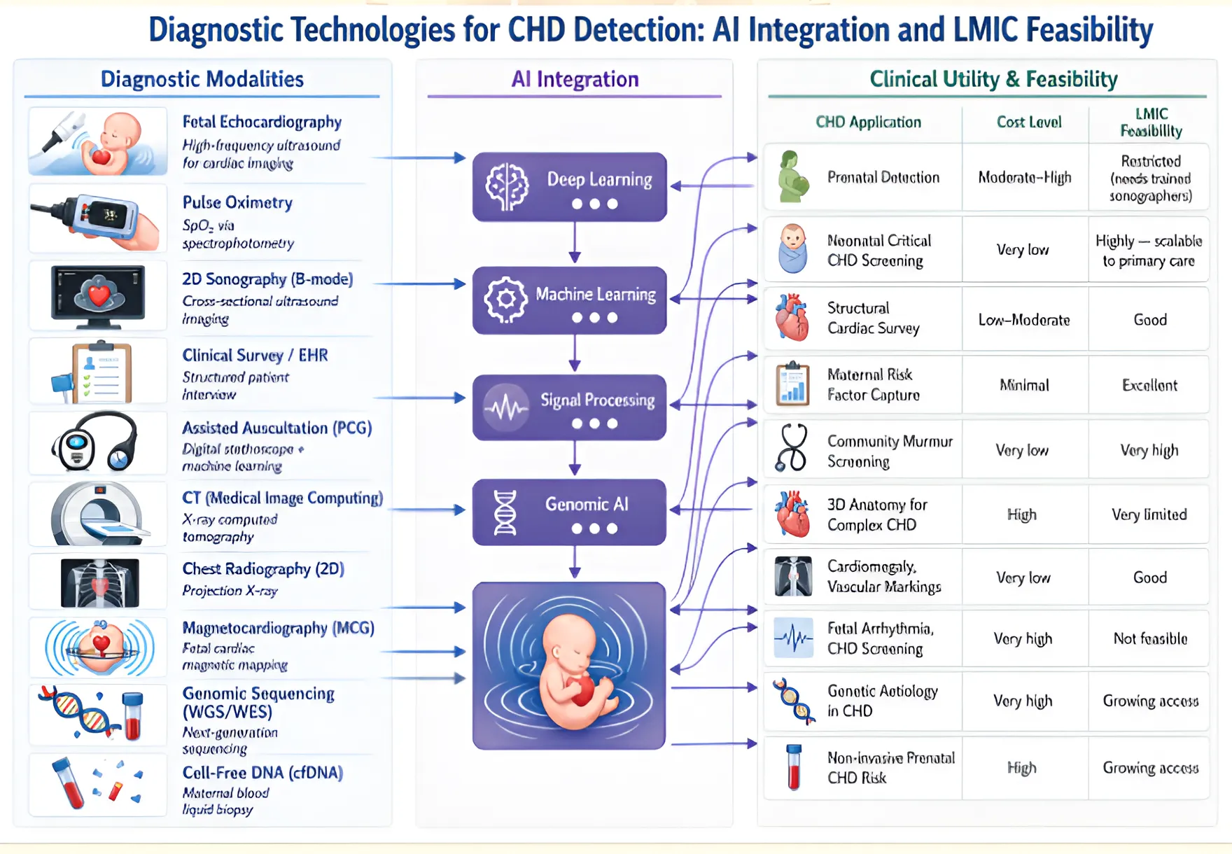
Figure 2 summarises a unified pipeline for CHD detection, linking diverse diagnostic modalities as pointed out in the above literature section, including imaging, physiological signals, clinical/EHR data, and genomics, corresponding AI techniques such as signal processing, machine learning, deep learning, and genomic AI, which transform raw data into predictive insights. Each method is useful in its own way, depending on the level of information the modality gives, cost incurred, and accessibility. Methods such as echocardiography and genomics provide precise information about structure or aetiology but require specialised resources and are costlier. Cheaper tools, such as pulse oximetry and clinical screening, can be used widely to screen large numbers of people and are widely recommended given their acceptable false-positive rate when established protocols are followed. By linking these methods to specific medical uses and to their practicality in low- and middle-income countries, the figure highlights an important balance between accuracy and ease of use.
Figure 2. The hierarchical mapping between diagnostic modalities and AI techniques, highlighting the trade-off between accessibility and diagnostic precision, particularly in LMIC settings
Diagnostic Technologies for CHD Detection: AI Integration and LMIC Feasibility
1.3 Risk Factors for Congenital Heart Disease
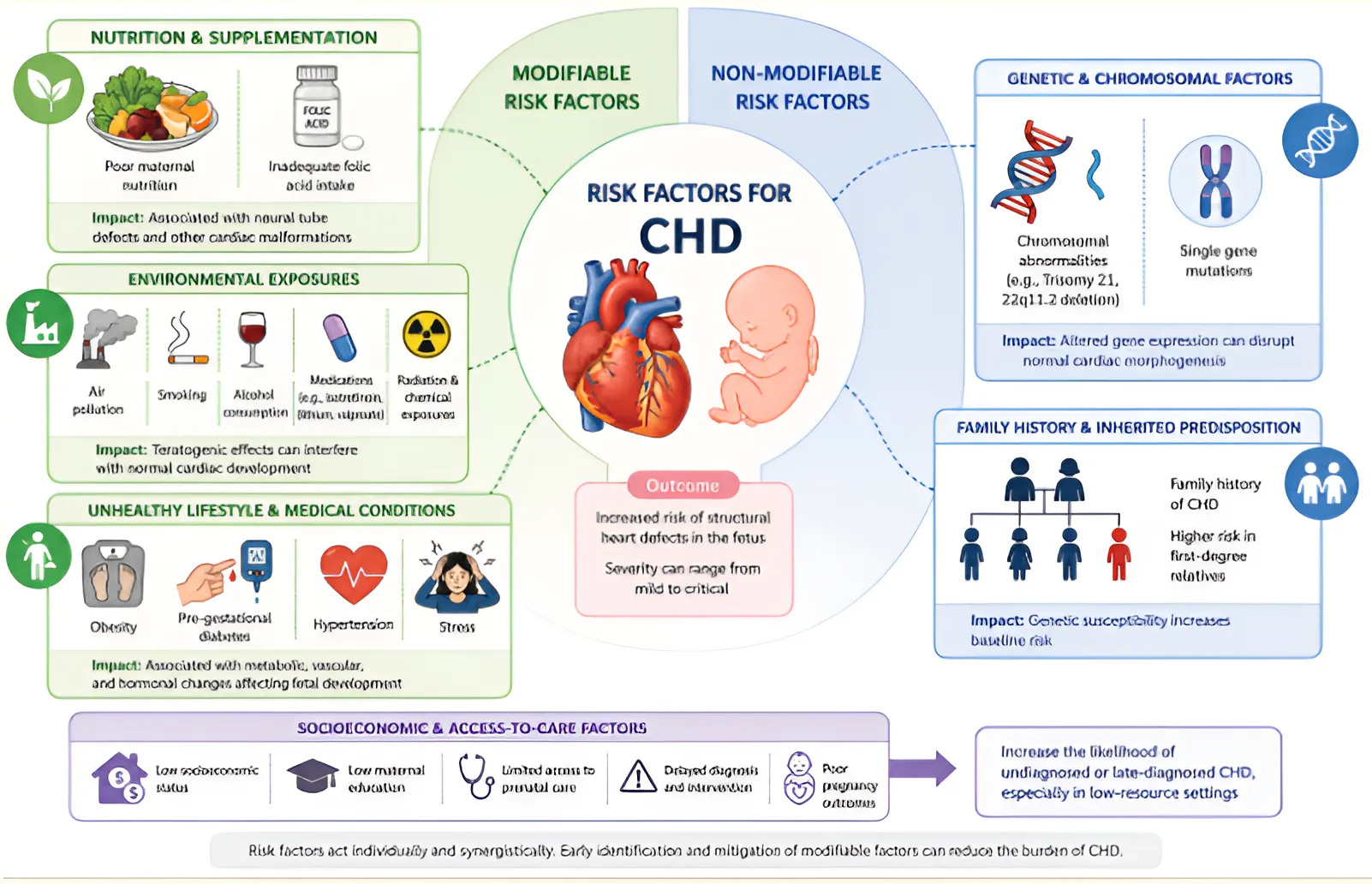
Around 25% of CHD's are classified as critical lesions that may become clinically apparent before the first scheduled neonatal examination. Its causes are multifactorial, arising from interacting genetic, maternal, environmental, and socioeconomic determinants. Identifying and quantifying these risk factors is important for both designing preventive strategies and building feature sets for ML prediction models. The Risk factors affecting CHD can be grouped into modifiable and nonmodifiable categories as shown in Figure 3. Non-genetic modifiable risk factors are attributes that,
when controlled, can reduce CHD risk and are therefore considered of prime importance. The modifiable risk factors include nutrition and folic acid supplementation, environmental exposures, and an unhealthy lifestyle. The attributes contributing to CHD are called Risk factors, and related risk factors are grouped under an indicator variable. Non-modifiable risk factors are characteristics that cannot be changed or controlled, but they increase the likelihood of CHD. These are mainly genetic and inherent factors, such as family history and genetic composition.
Figure 3. Maternal Risk Factors for CHD
CHD develops through a sequential and interconnected process spanning genetic predisposition, maternal risk exposure, and structural manifestation, as illustrated in the Figure 4. This multiscale progression underscores the limitations of isolated data modalities and indicates that CHD prediction should be viewed as a temporally evolving multimodal learning problem.
Figure 4. CHD Disease Detection pipeline
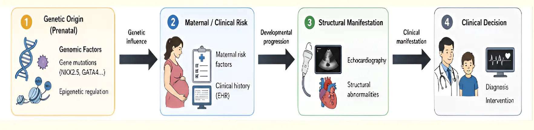
While diagnostic modalities enable the detection of structural abnormalities, they do not fully capture the upstream determinants of CHD risk. This necessitates a complementary understanding of underlying risk factors.
Family History: A positive family history, which includes consanguinity and affected relatives, suggests that genetic factors contribute to the condition. The risk of CHD recurrence among children who have affected parents or parents with past congenital anomalies in their family history shows a significant increase.
Sociodemographic and Maternal Demographic Factors: Low maternal educational attainment has been identified as an independent CHD risk factor. A meta-analysis of 64 observational studies, which included 182290 CHD cases, found no significant association with maternal age. The analysis showed that women who reached advanced maternal age (35 years or older) had a slightly increased risk, which was only detected in the adjusted estimates.
The income level of a family determines their socioeconomic status, which subsequently affects their access to proper nutrition and medical services, their exposure to different environmental factors, and their use of prenatal screening services.
Nutrition and Supplementation: Research shows that mothers-to-be who lack essential micronutrients, especially folic acid and iron, have a higher chance of CHD occurrence.
Environmental Exposure: The body can lose its ability to process environmental toxins from pollutants, radiation, or contaminated water during the first three months of pregnancy, which disrupts the normal process of organ development.
Unhealthy Lifestyle: Research has categorised alcohol use, tobacco use, area nut consumption, drug use, and passive smoking as modifiable behaviours that impact fetal oxygen supply and organ.
Maternal Medical Conditions: The maternal health conditions present before pregnancy, which include hypertension, thyroid disorders, anaemia, tuberculosis, psychiatric illness, cancer, prior miscarriages, and assisted conception, disrupt blood flow, metabolism, and immune function, create potential risks for fetal heart development. The strongest modifiable risk factor for CHD from the literature is pre-gestational diabetes mellitus, resulting in a fourfold risk. Maternal clotting disorders confer approximately ninefold excess CHD risk, with enoxaparin prescription in the first trimester also associated with elevated risk. Maternal obesity with BMI ≥ 30 is associated with a 29% excess CHD risk across 23 pooled studies, mediated by hyperinsulinaemia, hyperglycaemia, reduced folate status, and elevated systemic cytokines.
Medications Used during pregnancy: Fertility drugs, psychoactive medications, anaesthesia exposure, and insulin-dependent diabetes treatment were grouped due to their potential pharmacological and teratogenic effects.
Complications in Pregnancy: Psychological stress, accidents, and other pregnancy complications were grouped as acute systemic stressors that may indirectly influence fetal development through inflammatory or hemodynamic pathways.
2 Prediction of CHD Using EHR Data
This section presents the studies that applied machine learning to predict CHD using structured clinical tabular data. Various studies have shown that EHR-based machine learning methods can predict CHD, but their accuracy depends on the models used, how they handle data imbalance, and the size of their datasets. The study evaluated a three-layer backpropagation neural network (BPNN) on maternal clinical data and found that nonlinear systems outperformed logistic regression by capturing complex interactions among risk factors. The subsequent studies raised awareness of extreme class imbalance, and a study used cost-sensitive weighted SVM (WSVM) to achieve an AUC of 0.819 by penalising minority-class misclassifications. In the work the authors developed new cluster-then-classify methods that employed Balanced Random Forest and Easy-ensemble to improve recall by leveraging resampling and ensemble learning techniques. The researchers employed gradient boosting decision trees (GBDT) on Quebec claims and hospitalisation databases to conduct their research at a larger scale, which enabled tree-based ensembles to use high-dimensional ICD-coded data for automatic CHD detection. Optimisation was incorporated into the study by combining particle swarm optimisation (PSO) with a Support Vector Machine (SVM) to enhance hyperparameter tuning; however, the limitations were that the data used were from a single centre only. The meta-analysis on neural network ensembles showed that these systems deliver excellent performance across studies, yet there is insufficient validation specific to low- and middle-income countries. The clinical scoring system uses simple rules to produce results that researchers validated against echocardiography.
From a computational perspective, the risk factors used in the above studies form the foundational feature space for EHR-based predictive models. However, not all risk factors contribute equally to model performance. Strong predictors such as nutrition taken during pregnancy, pre-gestational diabetes, and genetic predisposition demonstrate high discriminative power. The results obtained from these models are difficult to generalise because the datasets contain an extremely low percentage of CHD cases. Risk-factor-based models cannot function as complete diagnostic systems because they only identify probabilistic associations between variables.
The predictive accuracy of machine learning models that use EHR data has high discriminative ability for CHD classification, with AUROC scores ranging between 0.82 and 0.99. EHR-based analyses identify statistical patterns rather than causal relationships. These approaches can support initial risk assessment but cannot directly identify structural heart defects that doctors need for diagnosing CHD. The EHR-based models aid in screening and triage processes, but they do not provide sufficient information for diagnosis.
The literature reviewed suggests the need to combine risk-based features with imaging and genomic data to improve predictive model performance. The next section details studies that used echocardiography modality. The Table 1 summarises the experimental details of the CHD prediction studies using EHR data.
1. Machine learning models for CHD prediction using structured clinical and EHR data
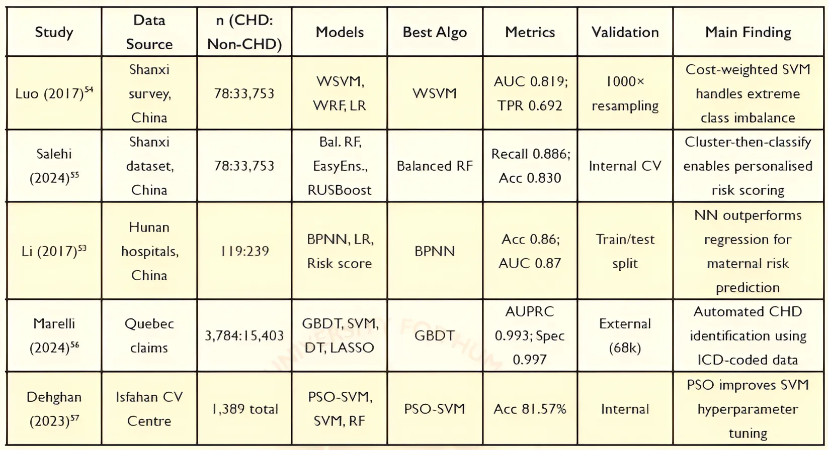
Study: Luo (2017) - Data Source: Shanxi survey, China - n (CHD: Non-CHD): 78:33,753 - Models: WSVM, WRF, LR - Best Algo: WSVM - Metrics: AUC 0.819; TPR 0.692 - Validation: 1000× resampling - Main Finding: Cost-weighted SVM handles extreme class imbalance
Study: Salehi (2024) - Data Source: Shanxi dataset, China - n: 78:33,753 - Models: Bal. RF, EasyEns., RUSBoost - Best Algo: Balanced RF - Metrics: Recall 0.886; Acc 0.830 - Validation: Internal CV - Main Finding: Cluster-then-classify enables personalised risk scoring
Study: Li (2017) - Data Source: Hunan hospitals, China - n: 119:239 - Models: BPNN, LR, Risk score - Best Algo: BPNN - Metrics: Acc 0.86; AUC 0.87 - Validation: Train/test split - Main Finding: NN outperforms regression for maternal risk prediction
Study: Marelli (2024) - Data Source: Quebec claims - n: 3,784:15,403 - Models: GBDT, SVM, DT, LASSO - Best Algo: GBDT - Metrics: AUPRC 0.993; Spec 0.997 - Validation: External (68k) - Main Finding: Automated CHD identification using ICD-coded data
Study: Dehghan (2023) - Data Source: Isfahan CV Centre - n: 1,389 total - Models: PSO-SVM, SVM, RF - Best Algo: PSO-SVM - Metrics: Acc 81.57% - Validation: Internal - Main Finding: PSO improves SVM hyperparameter tuning
3 Prediction of CHD Using Echocardiographic Data
Echocardiography has emerged as one of the most widely adopted modalities for CHD diagnosis, owing to its real-time imaging capability and high diagnostic value in identifying structural cardiac abnormalities.
3.1 Classic Machine Learning Approaches
Early automated approaches to CHD detection using echocardiography data relied on conventional machine learning with handcrafted features. The study applied a Random Forest (RF) classifier to 3,910 patients from a tertiary unit in Vietnam, one of the very few prenatal CHD AI studies conducted in a low- to-middle-income country setting, performing binary classification of fetal hearts using manually extracted cardiac biometric parameters. Similarly, SVM was used to extract textural features from 2D-echo videos to classify mitral regurgitation severity as normal, mild, moderate, or severe. These methods rely on manual feature engineering, which limits their ability to identify fine anatomical structures and subtle morphological differences in ultrasound images, particularly when the images are unclear. Because of these problems, researchers started using Deep Learning (DL) systems that can automatically find important features during the process.
3.2 Convolutional Neural Network (CNN)-Based Feature Extraction and Transfer Learning
Transfer learning (TL) has been used to apply deep CNNs pretrained on extensive natural image datasets to cardiac imaging tasks. It has become a leading method in echocardiographic analysis, using pretrained CNNs to capture overall visual features from ultrasound images. Figure 5 shows the comparison between essential CNN models that researchers employ in echocardiographic AI, showing their respective parameter counts and system depths and their ability to process ultrasound data. Recent studies indicate a shift from traditional transfer-learning approaches toward methods that support domain-specific representation learning.
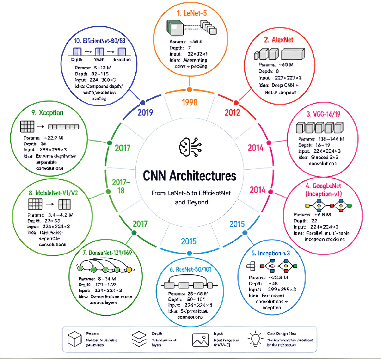
TL models are used for view classification,61 and the EchoNet-Dynamic system62 show that pretrained models can reach high diagnostic accuracy, with AUC scores between about 0.90 and 0.97. These methods save training time and work better, especially when there is limited labelled medical data.
CNNs have made echocardiographic analysis more effective by learning spatial patterns from raw data. Transfer learning, which leverages CNN weights trained on large datasets such as ImageNet, has been widely adopted in medical imaging because labelled echocardiographic datasets are often limited.63,64 AIEchoDx65 framework predicts heart disease subtypes from apical 4-chamber videos. The study66 used a multi-view CNN model on 1,308 patients to automatically diagnose ventricular septal defect (VSD) and atrial septal defect (ASD), reaching an accuracy of 93.9% by applying TL. A multi-view DL model trained on 1,128 case data and tested on 141, achieved 91% accuracy and an AUC of 0.92 for VSD and ASD diagnosis, also used TL.67 A multi-view multi-modal fusion framework68 was validated for 1,932 children, training 10 sub-models across five standard transthoracic echocardiography views and two imaging modalities, achieving a cross-centre AUC of 0.990 and the accuracy of clinicians with under three years’ experience improved from 0.707 to 0.953 with AI assistance. CHDNet,69 a Bayesian echo video classifier demonstrated promising predictive probability distributions.
In a different heart problem, applied a deep learning model to ultrasound data from 473 training and 277 test cases to detect coarctation of the aorta (CoA) in newborns, achieving a CoA error rate of 7.7% across 840 patients. In a study authors made use of CNN for identifying patent ductus arteriosus (80% accuracy) and in the study CNN was used for detecting ASD using colour doppler (AUC 0.92) and in CNN was used for 3D ultrasound segmentation in hypoplastic left heart syndrome (HLHS).
Some of the studies below used self-supervised learning harnessing CNNs. In the research, a self-supervised segmentation pipeline was developed that used watershed segmentation, Hough circle detection, and clinical shape priors to create weak labels, which were then improved through U-Net training on more than 18000 echocardiograms. The label-free pipeline achieved LV (Left Ventricle) Dice of 0.89 and r2 of 0.55–0.84 for chamber parameter estimation, with correlations with cardiac MRI comparable to those of supervised clinical echocardiography. The study by used an ensemble model modified U-Net to segment thoracic and cardiac structures from A4C images, while an ensemble classifier computed cardiothoracic ratio, cardiac axis, and fractional area change as transparent medical diagnostic intermediates. The first segmentation and EF estimation model specifically designed for pediatric use, EchoNet-Peds, achieved an MAE of 3.66% and an AUROC of 0.954 across the age range of 0 to 18 years, which surpassed the performance of the adult-trained EchoNet-Dynamic model on pediatric data (p < 10-100), thereby demonstrating that age-specific training data needs to be used for effective performance.
Direct classification methods provide quick results while needing fewer labels, but lack an understanding of anatomical structure. The supervised segmentation approach requires extensive annotations because it yields precise anatomical information across all body parts. SKGC (Semantic-Level Knowledge Guided Classification) framework was developed to address the limited availability of annotations using a two-stage framework that requires only a small number of segmentation masks.
Most existing DL methods for echocardiography focus on classifying individual frames, but this does not align with how doctors make diagnoses. In practice, diagnosis is made at the patient-level by looking at information from the whole video sequence. Therefore, there is a clear need for advanced temporal modelling techniques that can capture long-range dependencies and contextual relationships across echocardiographic sequences to better align with real-world clinical diagnosis.
3.3 Transformer-Based Modelling in Medical Imaging
Temporal modelling is important in echocardiography because heart function changes over time. AIEchoDx uses a CNN to combine information over time; CHDNet uses Bayesian methods to estimate uncertainty; EchoCLR applies contrastive learning to keep spatial and temporal information consistent. However, all these methods focus on short or limited time periods. Sequential data are handled by Recurrent Neural Networks (RNNs), which may also fail to capture complex long-term dynamics due to vanishing gradients and limited context retention.
Transformer architectures can mitigate these limitations by enabling global temporal attention across entire sequences, making them well-suited for modelling the full cardiac cycle, including correlations between spatially and temporally distant systolic and diastolic events. The self-attention mechanism used by the transformer enables a model to determine the importance of different elements in a sequence by dynamically assigning attention weights based on their contextual relevance.
Vaswani et al. presented architecture of transformers, which works by enabling full-sequence global attention to overcome the limitations of RNNs and is increasingly recognised as the preferred architecture for long-range temporal modelling in biomedical sequences, motivating their application to the full-sequence temporal aggregation task in the proposed study. Self-attention allows transformer models to overcome the constraints that restrict CNN performance. This method enables a sequence element to access all other elements simultaneously, which results in complete contextual understanding without losing distant information.
Recent hybrid CNN-Transformer frameworks, including CTransCNN, demonstrate the potential to combine local feature extraction with global temporal modelling by using the chest X-ray dataset.
The EchoCLR research study introduced the first self-supervised learning framework designed to capture both spatial and temporal information from echocardiography videos using SimCLR-based contrastive learning for the classification of CHD, such as ASD and AS. Using different videos from the same datapoint, by rearranging video frames, even with a small dataset. The system reached an AUROC of 0.818 for identifying severe aortic stenosis after training on only 1% of labelled data, much better than the 0.612 AUROC from usual transfer learning.
The recently proposed EchoFocus-CHD, extends the PanEcho architecture by incorporating a study-level transformer encoder to aggregate temporal and cross-view information, enabling prediction of 12 critical and 8 non-critical CHD lesions. PanEcho is a multi-view echocardiography framework designed to integrate information from multiple cardiac ultrasound views for CHD analysis. The training data consisted of 3.6 million videos from 58000 echocardiograms collected from various testing sites around the world. The system recorded an AUC score of 94%, but it struggled with external referral groups, achieving an AUC score of 77%. However, the observed performance degradation across external cohorts highlights the limitations of current multi-task architectures in handling domain variability and temporal inconsistency in echocardiographic data. Addressing these challenges requires more robust temporal representation learning and domain-invariant feature extraction, which motivates the approach proposed in this study. Other types of transformers proposed in the literature are vision transformers and Swin transformer. Table 2 provides a summary of experimental details of echocardiographic AI studies reviewed.
Table 2. Comprehensive Summary of Echo-Based Deep Learning Studies for CHD. EF: Ejection Fraction; LV: Left Ventricle; ASD: Atrial Septal Defect; VSD: Ventricular Septal Defect; HLHS: Hypoplastic Left Heart Syndrome; SSL: Self-Supervised Learning; A4C: Apical four-chamber; MAE: Mean Absolute Error; AUC: Area Under ROC curve; Acc: Accuracy
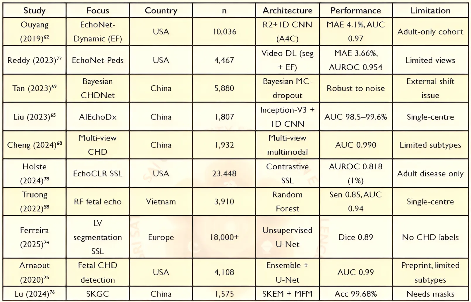
Study: Ouyang (2019) - Focus: EchoNet-Dynamic (EF) - Country: USA - n: 10,036 - Architecture: R2+1D CNN (A4C) - Performance: MAE 4.1%, AUC 0.97 - Limitation: Adult-only cohort
Study: Reddy (2023) - Focus: EchoNet-Peds - Country: USA - n: 4,467 - Architecture: Video DL (seg + EF) - Performance: MAE 3.66%, AUROC 0.954 - Limitation: Limited views
Study: Tan (2023) - Focus: Bayesian CHDNet - Country: China - n: 5,880 - Architecture: Bayesian MC-dropout - Performance: Robust to noise - Limitation: External shift issue
Study: Liu (2023) - Focus: AIEchoDx - Country: China - n: 1,807 - Architecture: Inception-V3 + ID CNN - Performance: AUC 98.5–99.6% - Limitation: Single-centre
Study: Cheng (2024) - Focus: Multi-view CHD - Country: China - n: 1,932 - Architecture: Multi-view multimodal - Performance: AUC 0.990 - Limitation: Limited subtypes
Study: Holste (2024) - Focus: EchoCLR SSL - Country: USA - n: 23,448 - Architecture: Contrastive SSL - Performance: AUROC 0.818 (1%) - Limitation: Adult disease only
Study: Truong (2022) - Focus: RF fetal echo - Country: Vietnam - n: 3,910 - Architecture: Random Forest - Performance: Sen 0.85, AUC 0.94 - Limitation: Single-centre
Study: Ferreira (2025) - Focus: LV segmentation SSL - Country: Europe - n: 18,000+ - Architecture: Unsupervised U-Net - Performance: Dice 0.89 - Limitation: No CHD labels
Study: Arnaout (2020) - Focus: Fetal CHD detection - Country: USA - n: 4,108 - Architecture: Ensemble + U-Net - Performance: AUC 0.99 - Limitation: Preprint, limited subtypes
Study: Lu (2024) - Focus: SKGC - Country: China - n: 1,575 - Architecture: SKEM + MFM - Performance: Acc 99.68% - Limitation: Needs masks
4 Prediction of CHD Using Genomic Data
Unlike imaging and EHR data, genomic information provides causal insight rather than observable phenotype, positioning it as a complementary modality rather than a standalone predictive tool.
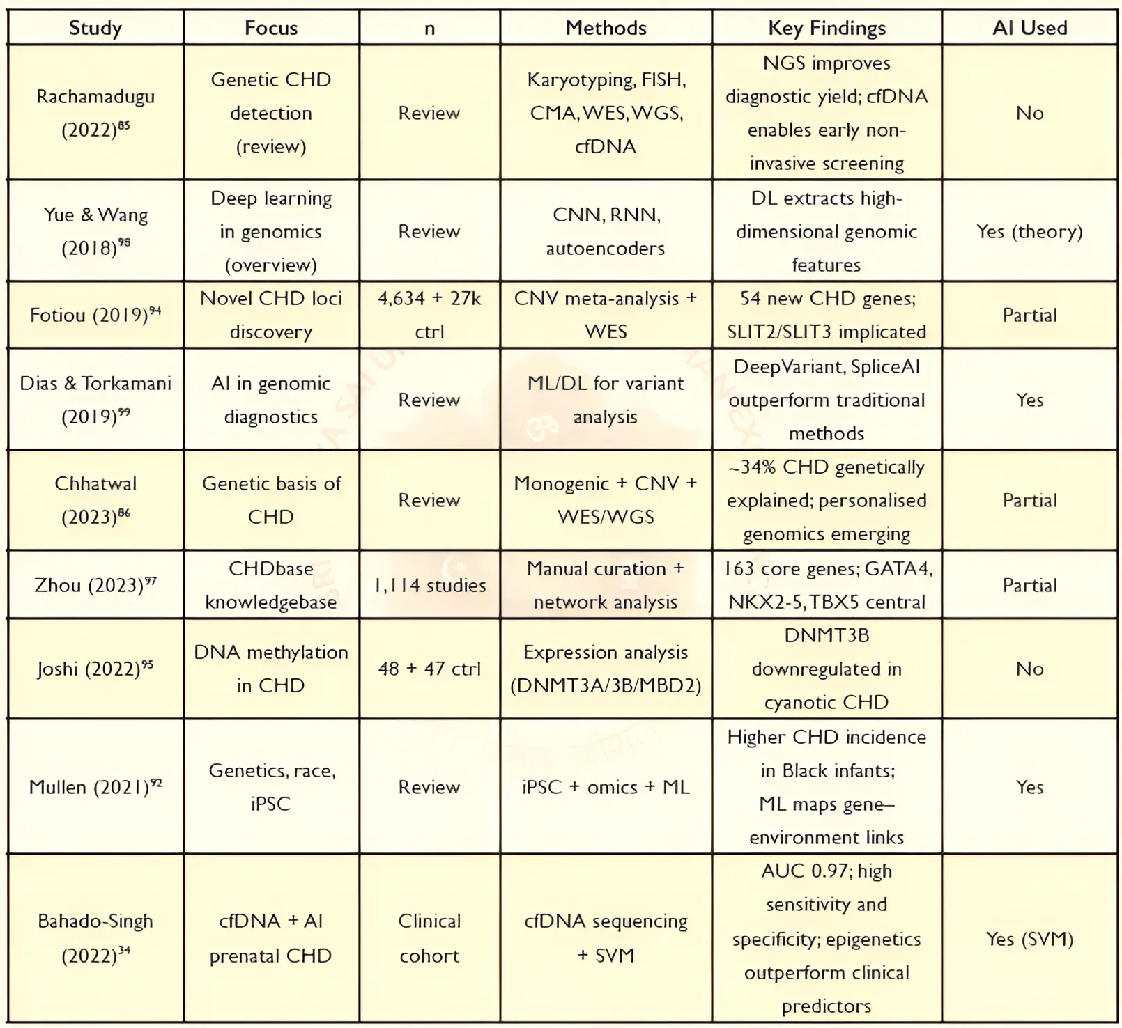
Genetic factors are estimated to explain 34 - 40% of CHD cases, encompassing chromosomal aneuploidies, copy number variants (CNVs), single-gene Mendelian disorders, and polygenic contributions. Sequencing technologies are getting widely used for accurate diagnosis for detecting genetic causes of CHD, the emerging role of AI in variant interpretation, non-invasive prenatal screening via cfDNA, and epigenomic methylation as a disease biomarker.
The diagnostic toolkit for genetic CHD spans multiple technologies. Karyotyping and fluorescence in situ hybridisation (FISH) identify chromosomal aneuploidies and major structural rearrangements; chromosomal microarray analysis (CMA) provides genome-wide CNV resolution; whole-exome sequencing (WES) targets protein-coding regions; and whole-genome sequencing (WGS) covers non-coding regulatory variants in addition to coding sequences.
Chromosomal aneuploidies principally trisomies 21 and 18 and monosomy X account for 8 - 10% of CHD cases; CNVs contribute a further 3 - 15%; and de novo variants (DNVs) identified from trio WES/WGS account for up to 20% of CHD cases associated with neurodevelopmental disorders. Approximately 400 genes have been linked to CHD, with cardiac transcription factors NKX2.5, GATA4, TBX1, and TBX5 occupying central positions in cardiogenic regulatory. iPSC-based patient-specific modelling combined with NGS, CRISPR editing, and ML offers a pathway to elucidating these race-related genetic contributors. Fotiou et al. demonstrated through metaanalysis of 4,634 nonsyndromic CHD cases 27,000+ controls that genes retained from ancestral whole-genome duplications known as ohnologs, are disproportionately represented among CHD candidate loci.
The process of cardiac embryogenesis (heart development in the fetus) depends on DNA methylation, which functions as a vital epigenetic control mechanism for gene expression. Disruption of methylation leads to the development of abnormal cardiac structures.
Zhou et al. assembled CHDbase, a curated knowledge base integrating 1,114 publications and linking 1,124 CHD-associated genes and 3,591 variants to over 300 CHD subtypes. Gene network analysis identified a core subnetwork of 163 highly connected genes, with GATA4, NKX2-5, and TBX5 having the highest centrality scores.
AI is reshaping genomic data interpretation. Deep learning models particularly CNNs and RNNs have demonstrated utility in variant calling, non-coding variant annotation, and phenotype-to-genotype mapping. CNN frameworks, such as DeepVariant, are used to detect SNVs and indels in sequencing reads, whereas SpliceAI identifies cryptic splice-site variants that account for at least 10% of rare pathogenic variation and are disproportionately relevant to CHD cases with unexplained aetiology. Phenotype-to-genotype mapping tools that integrate facial image analysis, NLP of clinical records, and deep-learning variant prioritization, exemplified by the PEDIA system, have been used to support automated diagnosis across hundreds of monogenic disorders simultaneously.
Circulating cfDNA in maternal blood, derived largely from the placenta, enables non-invasive access to fetal chromosomal and epigenomic information from early gestation. The study combined genome-wide epigenomic cfDNA analysis with a support vector machine (SVM) classifier for non-invasive fetal congenital heart disease (CHD) detection. The model achieved an AUC of 0.97, sensitivity of 98%, and specificity of 94%. The research also indicate that AI-powered cfDNA epigenomics as an innovative prenatal screening method that enables accurate assessment of CHD risk in the first trimester. Table 3 summarises the described genomic studies.
Table 3: Summary of genomic, epigenetic, and artificial intelligence-based approaches for CHD detection. NGS: next-generation sequencing; WES: whole-exome sequencing; WGS: whole-genome sequencing; CMA: chromosomal microarray analysis; cfDNA: cell-free DNA; CNV: copy number variant; SVM: support vector machine; DL: deep learning; iPSC: induced pluripotent stem cell.
Study: Rachamadugu (2022) - Focus: Genetic CHD detection (review) - n: Review - Methods: Karyotyping, FISH, CMA, WES, WGS, cfDNA - Key Findings: NGS improves diagnostic yield; cfDNA enables early non-invasive screening - AI Used: No
Study: Yue & Wang (2018) - Focus: Deep learning in genomics (overview) - n: Review - Methods: CNN, RNN, autoencoders - Key Findings: DL extracts high-dimensional genomic features - AI Used: Yes (theory)
Study: Fotiu (2019) - Focus: Novel CHD loci discovery - n: 4,634 + 27k ctrl - Methods: CNV meta-analysis + WES - Key Findings: 54 new CHD genes; SLIT2/SLIT3 implicated - AI Used: Partial
Study: Dias & Torkamani (2019) - Focus: AI in genomic diagnostics - n: Review - Methods: ML/DL for variant analysis - Key Findings: DeepVariant, SpliceAI outperform traditional methods - AI Used: Yes
Study: Chhatwal (2023) - Focus: Genetic basis of CHD - n: Review - Methods: Monogenic + CNV + WES/WGS - Key Findings: 34% CHD genetically explained; personalised genomics emerging - AI Used: Partial
Study: Zhou (2023) - Focus: CHDbase knowledgebase - n: 1,114 studies - Methods: Manual curation + network analysis - Key Findings: 163 core genes; GATA4, NKX2-5, TBX5 central - AI Used: Partial
Study: Joshi (2022) - Focus: DNA methylation in CHD - n: 48 + 47 ctrl - Methods: Expression analysis (DNMT3A/3B/MBD2) - Key Findings: DNMT3B downregulated in cyanotic CHD - AI Used: No
Study: Mullen (2021) - Focus: Genetics, race, iPSC - n: Review - Methods: iPSC + omics + ML - Key Findings: Higher CHD incidence in Black infants; ML maps gene–environment links - AI Used: Yes
Study: Bahado-Singh (2022) - Focus: cfDNA + AI prenatal CHD - n: Clinical cohort - Methods: cfDNA sequencing + SVM - Key Findings: AUC 0.97; high sensitivity and specificity; epigenetics outperform clinical predictors - AI Used: Yes (SVM)
5 Multimodal Integration
Multimodal data refers to the integration of information from multiple data sources or modalities, such as clinical records, medical imaging, genomic data, and physiological signals, to provide a more comprehensive understanding of disease. The absence of CHD-specific multimodal fusion studies represents the most significant gap in the field. Just as clinicians combine imaging findings, laboratory results, patient history, and clinical narratives to reach a diagnostic decision, computational models integrating complementary data sources may improve the robustness and generalisability of model predictions than their unimodal counterparts.
5.1 Fusion Strategies and Evidence
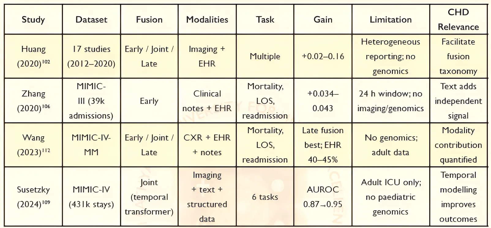
In a systematic review of 17 studies (2012-2020), a taxonomy of three multimodal fusion approaches was formalised. Early fusion (feature-level) concatenates extracted features from all modalities prior to model training, simple to implement but prone to dimensionality-related overfitting when high-dimensional imaging features are combined with sparse clinical data. Joint fusion (intermediate) trains modality-specific encoders simultaneously, which can promote complementary representation learning, at the cost of greater architectural complexity. Late fusion (decision-level) trains separate models per modality and aggregates their outputs, offering flexibility and robustness to missing data. Across all reviewed studies, multimodal models consistently outperformed unimodal baselines in accuracy by 1.2-27.7% and in AUROC by 0.02-0.16, with no single fusion strategy universally dominant.
The study operationalised multimodal fusion on the MIMIC-III critical care database using two architectures, Fusion-CNN and Fusion-LSTM, that combined static demographic variables, temporal vital signs and laboratory results. Similarly, the study added unstructured clinical notes that improved AUROC by 0.043 for Fusion-CNN and 0.034 for Fusion-LSTM on the in-hospital mortality task, confirming that clinical narratives encode prognostic information not represented in structured features alone.
The research extended the multimodal fusion to three modalities, including chest X-ray (CXR) images, structured EHR data, and radiology notes, on the MIMIC-IV-MM dataset. SHAP analysis (an explainable AI technique that interprets ML predictions by quantifying the contribution of each feature to the model's output) attributed approximately 40 - 45% of predictive contribution to structured EHR variables, with CXR contributing least for readmission prediction (where longitudinal clinical context dominates) and radiology notes providing intermediate value. Susetzky et al. introduced TAMME (Time-Aware MultiModal Transformer Encoder). Liang et al. introduced a unified architecture that treats the complete longitudinal EHR, including numerical, categorical, textual, and image data, as a single temporally ordered token sequence processed by a standard transformer encoder. BioLORD-2023 integrated semantic text representation with a knowledge graph. Table 4 summarises the experimental details of reviewed multimodal studies.
Table 4: Overview of multimodal artificial intelligence studies combining imaging, electronic health records, and clinical text for healthcare prediction tasks. Although none of the studies were conducted specifically for congenital heart disease (CHD), they demonstrate the potential of multimodal fusion strategies for improving predictive performance and provide a foundation for future CHD-specific multimodal diagnostic frameworks. EHR: electronic health record; CXR: chest X-ray; AUROC: area under the receiver operating characteristic curve; AUPRC: area under the precision-recall curve; LOS: length of stay.
Study: Huang (2020) - Dataset: 17 studies (2012–2020) - Fusion: Early / Joint / Late - Modalities: Imaging + EHR - Task: Multiple - Gain: +0.02–0.16 - Limitation: Heterogeneous reporting; no genomics - CHD Relevance: Facilitate fusion taxonomy
Study: Zhang (2020) - Dataset: MIMIC-III (39k admissions) - Fusion: Early - Modalities: Clinical notes + EHR - Task: Mortality, LOS, readmission - Gain: +0.034–0.043 - Limitation: 24 h window; no imaging/genomics - CHD Relevance: Text adds independent signal
Study: Wang (2023) - Dataset: MIMIC-IV MM - Fusion: Early / Joint / Late - Modalities: CXR + EHR + notes - Task: Mortality, LOS, readmission - Gain: Late fusion best; EHR 40–45% - Limitation: No genomics; adult data - CHD Relevance: Modality contribution quantified
Study: Susetzky (2024) - Dataset: MIMIC-IV (43k stays) - Fusion: Joint (temporal transformer) - Modalities: Imaging + text + structured data - Task: 6 tasks - Gain: AUROC 0.87–0.95 - Limitation: Adult ICU only; no paediatric genomics - CHD Relevance: Temporal modelling improves outcomes
5.2 Relevance to CHD and Research Gaps
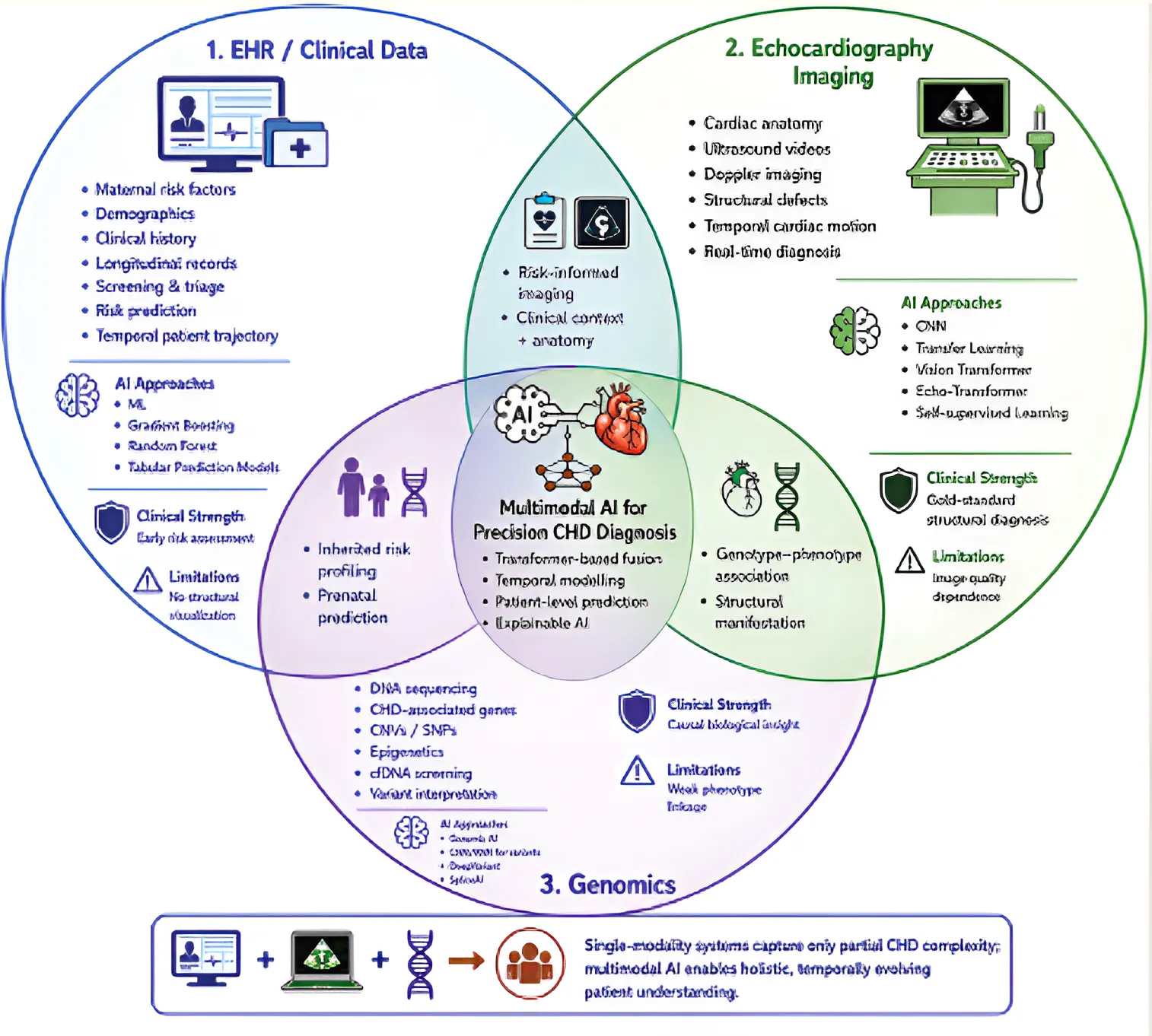
Multimodal integration is necessary because individual modalities have distinct limitations when considered in isolation for CHD prediction. The multi-factorial nature of CHD develops in three stages, which begin with genetic factors that impact heart development and continue through maternal and environmental influences on pregnancy risk and the final stage presentation of structural abnormalities through imaging. This complete trajectory is associated with multiple modalities to achieve full understanding. Multimodal approaches attempt to address this limitation by integrating heterogeneous data, enabling users to predict risks early and make precise assessments of structural problems. Many existing fusion methods have limited ability to capture temporal dependencies but it is essential for studying disease progression from prenatal to postnatal development.
Multimodal fusion studies targeted at CHD in paediatric populations are sparse. The current state of research shows that predictive systems based on AI can accurately predict CHD, but still have critical research gaps. Firstly, most studies analyse unimodal data, which limits their ability to understand how multiple factors contribute to coronary heart disease. Secondly, need to develop models that use time-dependent data to track patient progress from the prenatal period through postnatal development. Thirdly, paediatric-specific datasets, particularly from LMICs, are scarce, limiting the ability to apply research findings across diverse populations. Fourthly, there are few validation studies that reveal discrepancies between reported system performance and its actual use in real-world settings. The current state of explainability research limits its use in clinical settings due to its underdevelopment.
Addressing these gaps will likely require integrated multimodal systems that can process heterogeneous data types while still producing explainable and reliable results. CHD prediction inherently requires integrating precisely the modalities these architectures address: maternal demographic and social history (structured EHR), echocardiographic imaging, clinical documentation, and genomic variant data as compiled in the Figure 6.
Figure 6: Multimodal AI integration for CHD diagnosis
6 Proposed Framework
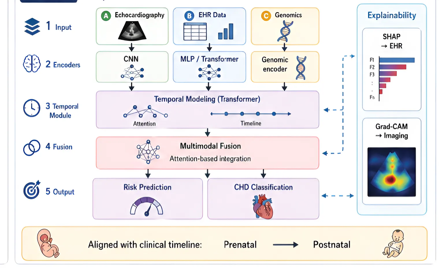
The gaps analysed lead to an approach to include a hierarchical multimodal framework, as shown in Figure 7, which uses multiple data sources from different time periods to improve CHD prediction accuracy. The model comprises three parts that process different types of data: CNN-based architectures handle echocardiographic imaging, neural networks process structured EHR data, and deep learning models analyse genomic features. The system uses sequence-based architectures, such as recurrent neural networks and transformers, to model temporal dependencies among patient data points. The proposed fusion module would use attention-based mechanisms to merge different representations, potentially allowing greater emphasis on clinically relevant features. The system
combines risk prediction and disease classification in its final output layer, while SHAP and Grad-CAM provide tools for developing understandable explanations. The framework integrates with existing clinical workflows to develop CHD prediction systems that deploy precision-based methods.
Figure 7: Proposed Fusion Model framework
7 Conclusions
The reviewed literature indicates that AI-based approaches have made substantial advances in CHD prediction, but the available methods still exhibit fragmented development and lack clinical effectiveness. EHR-based models detect initial health risks, echocardiographic tools provide precise assessments of structural defects through their diagnostic capabilities, and genomic techniques identify the genetic origins of diseases. However, no single approach captures the intricate tapestry of CHD.
The evidence suggests that predicting CHD is a layered, evolving challenge, with each data type offering unique insights at different moments in a patient's journey. Future systems must go beyond their current technical criteria to incorporate integrated systems that reflect actual medical practice. The transition to multimodal AI is more than a system upgrade because it could facilitate machines to make intelligent decisions and adapt to diverse contexts. The vision requires three essential actions: creating extensive multimodal databases for children, developing methods for ongoing assessment, and conducting thorough clinical testing of new technologies.
Moving from single-focus models to integrated, multimodal systems represent a pivotal step toward addressing the true complexity of CHD. The next wave of research should champion models that are not only explainable and validated in real-world clinical settings but also time-sensitive and
tailored to children and underserved regions. By closing the gap between technical breakthroughs and bedside care, multimodal AI could shift CHD detection from a game of catch-up to one of anticipation and prevention. The future of CHD prediction lies in weaving together diverse data sources into intelligent, time-aware systems that serve clinicians and patients alike.
Supplementary Materials: None.
Author Contributions: Shruthi S: Conceptualisation; Methodology; Investigation; Paper curation; Formal analysis; Writing - original draft, Visualisation, Validation, Project administration.
D Hanumanth Rao Naidu: Conceptualisation; Supervision; Writing - review and editing, Visualisation, Validation, Project administration.
Funding: This research received no external funding.
Institutional Review Board Statement: Not Applicable.
Informed Consent Statement: Not Applicable.
Data Availability Statement: Not Applicable.
Acknowledgements: Our heartfelt gratitude to Sadguru Sri Madhusudan Sai for this invaluable opportunity to contribute to this healthcare initiative, and to Sri Sathya Sai University for Human Excellence, Muddenahalli, Karnataka, for providing all essential resources required to conduct this research.
Conflicts of Interest: The author declares no conflicts of interest to report regarding the present study.
Declaration of Generative AI use: During the preparation of this work, the authors used ChatGPT (OpenAI, San Francisco, CA, USA) to assist in the preparation of the graphical abstract, figure layout design, and LaTeX table code generation. All AI-generated outputs were carefully reviewed, verified, edited, and validated by the authors to ensure scientific accuracy, completeness, and consistency with the study findings. The authors take full responsibility for the content of the published article.
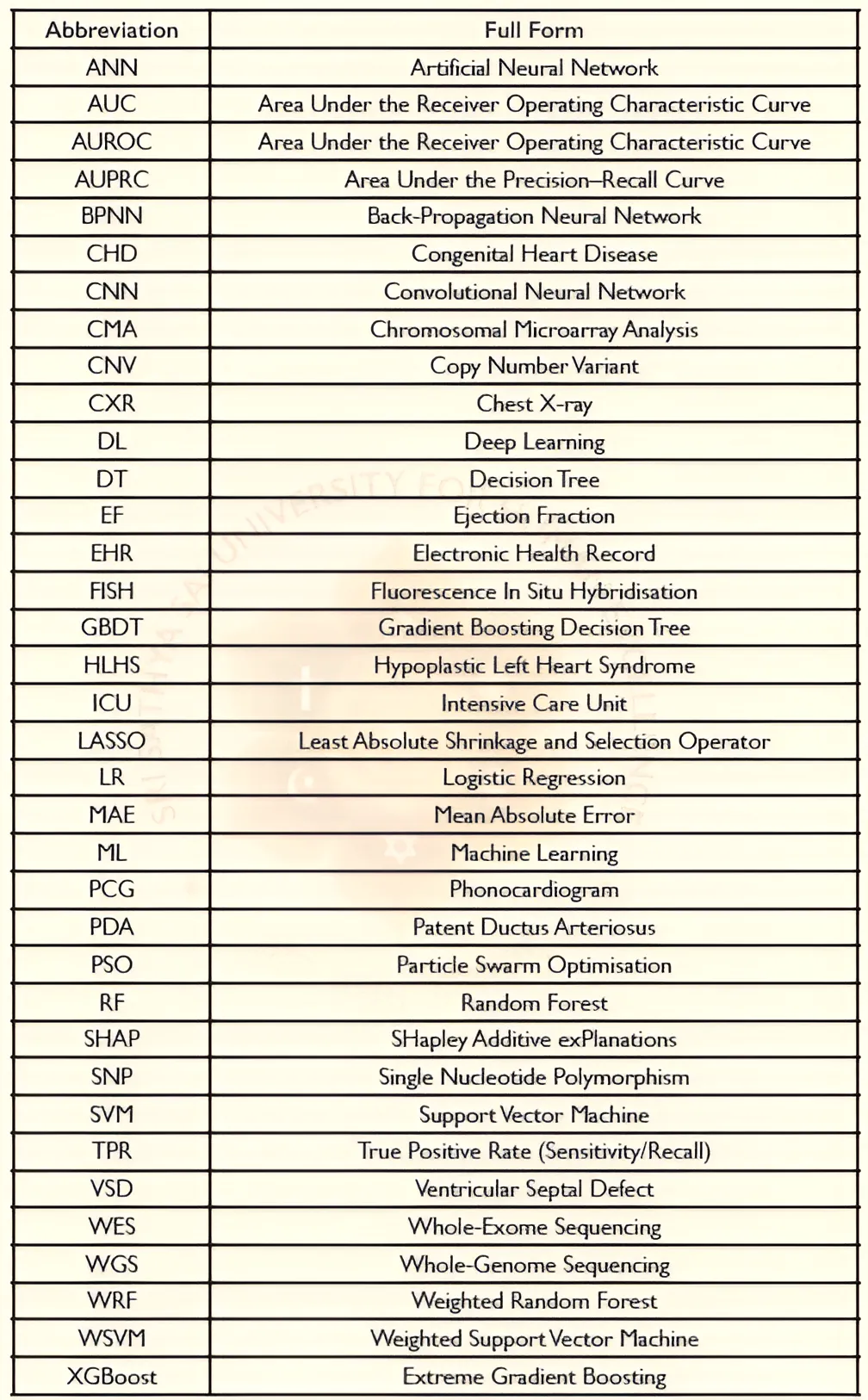
Abbreviations
-
J Xu, Q Li, L Deng, J Xiong, Z Cheng, and C Ye. Global, regional, and national epidemiology of congenital heart disease in children from 1990-2021. Frontiers in Cardiovascular Medicine, 12:1522644, 2025. doi:10.3389/fcvm.2025.1522644.
-
D van der Linde, E E M Konings, M A Slager, M Witsenburg, W A Helbing, J J M Takkenberg, and J W Roos-Hesselink. Birth prevalence of congenital heart disease worldwide: a systematic review and meta-analysis. Journal of the American College of Cardiology, 58(21):2241-2247, 2011. doi:10.1016/j.jacc.2011.08.025.
-
B J Bouma and B J M Mulder. Changing landscape of congenital heart disease. Circulation Research, 120(6):908-922, 2017. doi:10.1161/CIRCRESAHA.116.309302.
-
K S Iyer and S Sivalingam. Congenital heart disease in low- and middle-income countries: can india show the way? Indian Journal of Thoracic and Cardiovascular Surgery, 41(6):657-663, 2025. doi:10.1007/s12055-025-01969-w.
-
M E Pierpont, M Brueckner, W K Chung, V Garg, R V Lacro, A L McGuire, S Mital, J R Priest, W T Pu, A Roberts, S M Ware, B D Gelb, and M W Russell. Genetic basis for congenital heart disease: revisited: a scientific statement from the american heart association. Circulation, 138(21):e653-e711, 2018. doi:10.1161/CIR.0000000000000606.
-
A Saxena. Congenital heart disease in india: a status report. Indian Pediatrics, 55(12):1075-1082, 2018.
-
J I E Hoffman. The global burden of congenital heart disease. Cardiovascular Journal of Africa, 24(4):141-145, 2013. doi:10.5830/CVJA-2013-028.
-
L Deng, Q Li, and Z Cheng. Evaluating the global, regional, and national burden of congenital heart disease in infants younger than 1 year: a 1990-2021 systematic analysis for the gbd study 2021. Frontiers in Pediatrics, 13:1467914, 2025. doi:10.3389/fped.2025.1467914.
-
W Wu, J He, and X Shao. Incidence and mortality trend of congenital heart disease at the global, regional, and national level, 1990-2017. Medicine (Baltimore), 99(23):e20593, 2020. doi:10.1097/MD.00000000000020593.
-
D Vervoort, B Meuris, B Meyns, and P Verbrugge. Global cardiac surgery: access to cardiac surgical care around the world. Journal of Thoracic and Cardiovascular Surgery, 159(3):987-996, 2020. doi:10.1016/j.jtcvs.2019.04.039.
-
J I E Hoffman and S Kaplan. The incidence of congenital heart disease. Journal of the American College of Cardiology, 39(12):1890-1900, 2002. doi:10.1016/S0735-1097(02)01886-7.
-
F Bailliard and R H Anderson. Tetralogy of fallot. Orphanet Journal of Rare Diseases, 4:2, 2009. doi:10.1186/1750-1172-4-2.
-
S Ramegowda and N B Ramachandra. Parental consanguinity increases congenital heart diseases in south india. Annals of Human Biology, 33(5-6):519-528, 2006. doi:10.1080/03014460600909349.
-
D Sasikumar, M A Prabhu, R Kurup, E Francis, S Kumar, S T Gangadharan, K K Mahadevan, S Sivasankaran, and R K Kumar. Outcomes of neonatal critical congenital heart disease: results of a prospective registry-based study from south india. Archives of Disease in Childhood, 108:889-894, 2023. doi:10.1136/archdischild-2023-325471.
-
P K Jain, M Lazarus, A Tiwari, and V K Athwani. Prevalence and pattern of congenital heart disease in pediatric population—a study from central india. International Journal of Recent Surgical and Medical Sciences, 9:39-44, 2023. doi:10.1055/s-0042-1751085.
-
N Mishra, K Vagha, S Sawhney, S Murhekar, J D Vagha, and C K Javaji. Estimation of prevalence of thrombocytopenia in cyanotic congenital heart disease: a cross-sectional study among the pediatric population. Cureus, 16:e55453, 2024. doi:10.7759/cureus.55453.
-
R Bhardwaj, S K Rai, A K Yadav, S Lakhotia, D Agrawal, A Kumar, and B Mohapatra. Epidemiology of congenital heart disease in india. Congenital Heart Disease, 10:437-446, 2015.
-
Mehul Chaudhary, Anil J. Katara, Himanshu Gopat, and Yogesh Modi. A study of clinical profile morbidities and mortalities of congenital heart disease. Research Journal of Medical Sciences, 19(1):297-303, 2025. doi:10.36478/makrjms.2025.1.297.303.
-
R Singh and N Rajaram Tawker. The spectrum of congenital heart disease in children in the andaman and nicobar islands: a five-year retrospective study. Cureus, 14:e29109, 2022. doi:10.7759/cureus.29109.
-
R Amroliwala, M Rasania, R Memon, A Pokharna, and D Shah. Study of congenital heart diseases in neonates. International Journal of Contemporary Pediatrics, 5(1):75-81, 2018. doi:10.18203/2349-3291.ijcp20175541.
-
V K Ravilala, S Kotla, T Radhakishan, and R Malava. Study of congenital heart disease in neonates: clinical profile, diagnosis, immediate outcome and short-term follow-up. International Journal of Contemporary Pediatrics, 5(4):1304-1309, 2018. doi:10.18203/2349-3291.ijcp20182433.
-
K Doddabasava, P Philip, S B Shetty, C S Jacob, and K Subramanyam. Gamut of congenital heart diseases in a tertiary center in south india: an ode to echocardiography. International Journal of Contemporary Pediatrics, 4(3):1021-1026, 2017. doi:10.18203/2349-3291.ijcp20171720.
-
S Naik, M Irshad, A Kachroo, and M Ahmad. A study of prevalence and pattern of congenital heart disease at sopore, kashmir, north india. International Journal of Contemporary Pediatrics, 6(2):275-279, 2019. doi:10.18203/2349-3291.ijcp20190226.
-
A Kumar and P Kumar. Echocardiographic study of ventricular septal defect in 1- to 12-year-old children visiting a tertiary care center in patna, india. Cureus, 15:e46363, 2023. doi:10.7759/cureus.46363.
-
S Kishore, M Kumar, A Kumar, A Gupta, C Chandan, A Anshuman, J Prakash, S Sinha, and N Kumar. Clinical and echocardiographic profile of congenital heart diseases in the 0-12-year age group in a tertiary care medical institute in eastern india: a retrospective cross-sectional study. Cureus, 14:e26114, 2022. doi:10.7759/cureus.26114.
-
S M Nair, B Zheleva, A Dobrzycka, P Hesselin, R Sadanandan, and R K Kumar. A population health approach to address the burden of congenital heart disease in kerala, india. Global Heart, 16(1):71, 2021. doi:10.5334/gh.1034.
-
N K Jat, D K Bhagwani, N Bhutani, U Sharma, R Sharma, and R Gupta. Assessment of the prevalence of congenital heart disease in children with pneumonia in tertiary care hospital: a cross-sectional study. Annals of Medicine and Surgery, 73:103111, 2022. doi:10.1016/j.amsu.2021.103111.
-
Samuel Menahem, Arvind Sehgal, and Simon Meagher. Early detection of significant congenital heart disease: The contribution of fetal cardiac ultrasound and newborn pulse oximetry screening. Journal of Paediatrics and Child Health, 57(3):323-327, 2021. doi:10.1111/jpc.15355.
-
L. J. Salomon, Z. Alfirevic, V. Berghella, C. Bilardo, E. Hernandez-Andrade, S. L. Johnsen, K. Kalache, K.-Y. Leung, G. Malinger, H. Munoz, F. Prefumo, A. Toi, and W. Lee. Practice guidelines for performance of the routine mid-trimester fetal ultrasound scan. Ultrasound in Obstetrics & Gynecology, 37(1):116-126, 2011. doi:10.1002/uog.8831.
-
Zahra Hoodbhoy, Uswa Jiwani, Saima Sattar, Rehana Salam, Babar Hasan, and Jai K. Das. Diagnostic accuracy of machine learning models to identify congenital heart disease: A meta-analysis. Frontiers in Artificial Intelligence, 4:708365, 2021. doi:10.3389/frai.2021.708365.
-
John S. Chorba, Avi M. Shapiro, Le Le, John Maidens, John Prince, Steve Pham, Mia M. Kanzawa, Daniel N. Barbosa, Caroline Currie, Catherine Brooks, Brent E. White, Anna Huskin, Jason Paek, Jack Geocaris, Dinatu Elnathan, Ria Ronquillo, Roy Kim, Zenith H. Alam, Vaikom S. Mahadevan, Sophie G. Fuller, Grant W. Stalker, Sara A. Bravo, Dina Jean, John J. Lee, Medeona Gjergjindreaj, Christos G. Mihos, Steven T. Forman, Subramanian Venkatraman, Patrick M. McCarthy, and James D. Thomas. Deep learning algorithm for automated cardiac murmur detection via a digital stethoscope platform. Journal of the American Heart Association, 10(9):e019905, 2021. doi:10.1161/JAHA.120.019905.
-
Shi-Joon Yoo, Omar Thabit, Eul Kyung Kim, Haruki Ide, Deane Yim, Anreea Dragulescu, Mike Seed, Lars Grosse-Wortmann, and Glen Van Arsdell. 3d printing in medicine of congenital heart diseases. 3D Printing in Medicine, 2(1):3, 2016. doi:10.1186/s41205-016-0004-x.
-
Massimo Chessa, Alexander Van De Bruaene, Kanwal Farooqi, Israel Valverde, Christian Jung, Emiliano Votta, Francesco Sturla, Gerhard Paul Diller, Margarita Brida, Zhonghua Sun, Stephen H. Little, and Michael A. Gatzoulis. Three-dimensional printing, holograms, computational modelling, and artificial intelligence for adult congenital heart disease care: an exciting future. European Heart Journal, 43(28):2672-2684, 2022. doi:10.1093/eurheartj/eha266.
-
R Bahado-Singh, P Friedman, C Talbot, B Aydas, S Southekal, N K Mishra, et al. Cell-free dna in maternal blood and ai: prenatal detection of fetal chd. American Journal of Obstetrics and Gynecology, 2022. doi:10.1016/j.ajog.2022.07.062.
-
J. Espinoza, J. Caradeux, A. Bhimraj, K. H. Nicolaides, and M. Bennasar. Cell-free fetal dna for non-invasive prenatal testing: a review. Fetal Diagnosis and Therapy, 46(3):183-196, 2019. doi:10.1159/000497299.
-
Fazil M. Izhar, Shaad Abqari, Tabassum Shahab, and Syed Manazir Ali. Clinical score to detect congenital heart defects: Concept of second screening. Annals of Pediatric Cardiology, 13(4):281-288, 2020. doi:10.4103/apc.APC_113_19.
-
Matthew E. Oster, Matthew J. Strickland, and William T. Mahle. Racial and ethnic disparities in postoperative mortality following congenital heart surgery. The Journal of Pediatrics, 159(2):222-226, 2011. doi:10.1016/j.jpeds.2011.02.041.
-
L. Wu, N. Li, and Y. Liu. Association between maternal factors and risk of congenital heart disease in offspring: A systematic review and meta-analysis. Maternal and Child Health Journal, 27:29-48, 2023. doi:10.1007/s10995-022-03538-8.
-
S. Ramegowda and N.B. Ramachandra. Parental consanguinity increases congenital heart diseases in south india. Annals of Human Biology, 33(5-6):519-528, 2006. doi:10.1080/03014460600909349.
-
D. Dev, R. Sharma, M. Sharma, K. Agrawal, and M. Garg. Evaluation of consanguinity as a risk factor for congenital heart diseases. International Journal of Contemporary Pediatrics, 3(3):868-871, 2016. doi:10.18203/2349-3291.ijcp20162356.
-
H. Dolk, N. McCullough, S. Callaghan, F. Casey, B. Craig, J. Given, et al. Risk factors for congenital heart disease: The baby hearts study, a population-based case-control study. PLOS ONE, 15(2):e0227908, 2020. doi:10.1371/journal.pone.0227908.
-
S.T. Hashim, R.A. Alamri, R. Bakraa, R. Rawas, F. Farahat, and R. Waggass. The association between maternal age and the prevalence of congenital heart disease in newborns from 2016 to 2018 in a single cardiac center in jeddah, saudi arabia. Cureus, 12:e7463, 2020.
-
Q. Miao, S. Dunn, S.W. Wen, J. Lougheed, P. Yang, M. Davies, et al. Association between maternal marginalization and infants born with congenital heart disease in ontario, canada. BMC Public Health, 23:790, 2023. doi:10.1186/s12889-023-15660-5.
-
S. Santander Ballestin, M. I. Gimenez Campos, J. Ballestin Ballestin, and M. J. Luesma Bartolome. Is supplementation with micronutrients still necessary during pregnancy? a review. Nutrients, 13:3134, 2021. doi:10.3390/nu13093134.
-
R. Joshi, M. Bhoite, P. Mandhare, S. Nath, S. Kapoor, R. Wadke, et al. Population-based comparative nutritional status of unoperated congenital heart defects patients from a tertiary pediatric cardiac centre in india. 2025. doi:10.21203/rs.3.rs-5684483/v1.
-
R.O. Joshi, P. Kukshal, A. Kumar, P.R. Murthy, S. Chellappan, K. Manohar, et al. Congenital heart defects and environmental factors: A snapshot of chd cohort from a tertiary cardiac care centre in india. Annals of Pediatrics, 6(2):1123, 2023.
-
T. Wang, L. Chen, B. Ni, X. Sheng, P. Huang, S. Zhang, et al. Maternal pre-pregnancy/early-pregnancy smoking and risk of congenital heart diseases in offspring: A prospective cohort study in central china. Journal of Global Health, 12:11009, 2022. doi:10.7189/jogh.12.11009.
-
M. Alhindal, J. Janahi, E.C. D'Angelo, V. Lisignoli, R. Palmieri, A. Cutri, et al. Impact of smoking on cardiovascular health: Mechanisms, epidemiology and specific concerns regarding congenital heart disease. International Journal of Cardiology: Congenital Heart Disease, 20:100581, 2025. doi:10.1016/j.ijjchd.2025.100581.
-
C. Deng, J. Pu, Y. Deng, L. Xie, L. Liu, X. Guo, et al. Association between maternal smoke exposure and congenital heart defects from a case-control study in china. Scientific Reports, 12:14973, 2022. doi:10.1038/s41598-022-18909-y.
-
E. Helle and J. R. Priest. Maternal obesity and diabetes mellitus as risk factors for congenital heart disease in the offspring. Journal of the American Heart Association, 9:e011541, 2020. doi:10.1161/JAHA.119.011541.
-
S. Liu, K. S. Joseph, S. Lisonkova, J. Rouleau, M. Van den Hof, R. Sauve, et al. Association between maternal chronic conditions and congenital heart defects: A population-based study. Circulation, 128:583-589, 2013. doi:10.1161/circulationaha.112.001054.
-
H. Su, E. Guo, M. Woodward, J. R. He, T. Waterboer, A. Schuermans, et al. First trimester maternal infections and offspring congenital heart defects: A meta-analysis. European Heart Journal, 47:794-812, 2026. doi:10.1093/eurheartj/eha564.
-
Huixia Li, Miyang Luo, Jianfei Zheng, Jiayou Luo, Rong Zeng, Na Feng, Qiyun Du, and Junqun Fang. An artificial neural network prediction model of congenital heart disease based on risk factors. Medicine (Baltimore), 96(6):e6090, 2017. doi:10.1097/MD.0000000000006090.
-
Yanhong Luo, Zhi Li, Husheng Guo, Hongyan Cao, Chunying Song, Xingping Guo, and Yanbo Zhang. Predicting congenital heart defects: A comparison of three data mining methods. PLOS ONE, 12(5):e0177811, 2017. doi:10.1371/journal.pone.0177811.
-
Amirreza Salehi and Majid Khedmati. Identifying at-risk patients for congenital heart disease using integrated predictive models and fuzzy clustering analysis: A cross-sectional study. Heliyon, 10(21):e39609, 2024. doi:10.1016/j.heliyon.2024.e39609.
-
Ariane J. Marelli, C. Li, A. Liu, H. Nguyen, H. Moroz, James M. Brophy, et al. Machine learning informed diagnosis for congenital heart disease in large claims data. JACC Advances, 3(2):100801, 2024. doi:10.1016/j.jacadv.2023.100801.
-
B. Dehghan, M. R. Sabri, A. Ahmadi, M. Ghaderian, C. Mahdavi, D. Ramezani Nejad, et al. Identifying factors affecting chd incidence using svm and particle swarm optimisation. Advanced Biomedical Research, 12:130, 2023. doi:10.4103/abrab_231_22.
-
Vien T. Truong, Binh P. Nguyen, Thanh-Hoang Nguyen-Vo, Wojciech Mazur, Eugene S. Chung, Cassady Palmer, et al. Application of machine learning in screening for congenital heart diseases using fetal echocardiography. International Journal of Cardiovascular Imaging, 38(5):1007-1015, 2022. doi:10.1007/s10554-022-02566-3.
-
H. Moghaddasi and S. Nourian. Automatic assessment of mitral regurgitation severity using texture-based features and support vector machine. Computers in Biology and Medicine, 75:47-55, 2016. doi:10.1016/j.compbiomed.2016.05.009.
-
G. Huang, Z. Liu, L. Van Der Maaten, and K. Q. Weinberger. Densely connected convolutional networks. In Proceedings of the IEEE Conference on Computer Vision and Pattern Recognition (CVPR), pages 4700-4708, 2017. doi:10.1109/CVPR.2017.243.
-
Ali Madani, Ramy Arnaout, Mohammad Mofrad, and Rima Arnaout. Fast and accurate view classification of echocardiograms using deep learning. npj Digital Medicine, 1:6, 2018. doi:10.1038/s41746-017-0013-1.
-
David Ouyang, Bryan He, Amirata Ghorbani, Neal Yuan, Joseph Ebinger, Curtis P. Langlotz, et al. Video-based ai for beat-to-beat assessment of cardiac function. Nature, 580(7802):252-256, 2020. doi:10.1038/s41586-020-2145-8.
-
M. Tan and Q. Le. EfficientNet: Rethinking model scaling for convolutional neural networks. In Proceedings of the IEEE/CVF Conference on Computer Vision and Pattern Recognition (CVPR), pages 6105-6114, 2019. doi:10.48550/arXiv.1905.11946.
-
K. He, X. Zhang, S. Ren, and J. Sun. Deep residual learning for image recognition. In Proceedings of the IEEE Conference on Computer Vision and Pattern Recognition (CVPR), pages 770-778, 2016. doi:10.1109/CVPR.2016.90.
-
B. Liu, H. Chang, D. Yang, F. Yang, Q. Wang, Y. Deng, et al. A deep learning framework assisted echocardiography with diagnosis, lesion localization, phenogrouping heterogeneous disease, and anomaly detection. Scientific Reports, 13:3, 2023. doi:10.1038/s41598-022-27211-w.
-
S. Wang et al. Multi-view convolutional neural network for diagnosis of ventricular septal defect and atrial septal defect. IEEE Transactions on Medical Imaging, 39, 2020. doi:10.1109/TMI.2020.3046010.
-
X. Jiang, J. Yu, J. Ye, W. Jia, W. Xu, and Q. Shu. A deep learning-based method for pediatric congenital heart disease detection with seven standard views in echocardiography. World Journal for Pediatric Surgery, 6:e000580, 2023. doi:10.1136/wjps-2023-000580.
-
M. Cheng, J. Wang, X. Liu, Y. Wang, Q. Wu, F. Wang, et al. Development and validation of a deep-learning network for detecting congenital heart disease from multi-view multi-modal transthoracic echocardiograms. Research, 7:0319, 2024. doi:10.34133/research.0319.
-
J. Tan et al. CHDNet: Bayesian deep learning for congenital heart disease diagnosis with uncertainty estimation. IEEE Journal of Biomedical and Health Informatics, 26, 2022. doi:10.1016/j.eng.2022.10.015.
-
F. Pereira, A. Bueno, A. Rodriguez, D. Perrin, G. Marx, M. Cardinale, et al. Automated detection of coarctation of aorta in neonates from two-dimensional echocardiograms. Journal of Medical Imaging, 4(1):014502, 2017. doi:10.1117/1.JMI.4.1.014502.
-
Jason Erno, Thomas Gomes, Christopher Baltimore, John P. Lineberger, D. Hudson Smith, et al. Automated identification of patent ducts arteriosus using a computer vision model. Journal of Ultrasound in Medicine, 2023. doi:10.1002/jum.16305.
-
X. Lin, F. Yang, Y. Chen, X. Chen, W. Wang, W. Li, et al. Echocardiography-based ai for detection and quantification of atrial septal defect. Frontiers in Cardiovascular Medicine, 10:985657, 2023. doi:10.3389/fcvm.2023.985657.
-
Christian Herz, Danielle F. Pace, and et al. Nam. Segmentation of tricuspid valve leaflets from transthoracic 3d echocardiograms of children with hypoplastic left heart syndrome using deep learning. Frontiers in Cardiovascular Medicine, 2021. doi:10.3389/fcvm.2021.735587.
-
D. L. Ferreira, C. Lau, Z. Salaymang, and R. Arnaout. Self-supervised learning for label-free segmentation in cardiac ultrasound. Nature Communications, 16:4070, 2025. doi:10.1038/s41467-025-4070-0.
-
R. Arnaout, L. Curran, Y. Zhao, J.C. Levine, E. Chinn, and A.J. Moon-Grady. Expert-level prenatal detection of complex congenital heart disease from screening ultrasound. medRxiv, 2020. doi:10.1101/2020.06.22.20137786.
-
Y. Lu, G. Tan, B. Pu, H. Wang, B. Liang, K. Li, et al. Skgc: Semantic-level knowledge-guided classification for fetal congenital heart disease. IEEE Journal of Biomedical and Health Informatics, 28(10):6105-6116, 2024. doi:10.1109/JBHI.2024.3381234.
-
C. D. Reddy, L. Lopez, D. Ouyang, J. Y. Zou, and B. He. Video-based deep learning for left ventricular ejection fraction in pediatric patients. Journal of the American Society of Echocardiography, 36(5):482-489, 2023. doi:10.1016/j.echo.2022.12.014.
-
G. Holste, E. K. Oikonomou, B. J. Mortazavi, Z. Wang, and R. Khera. Self-supervised learning of echocardiogram videos for aortic stenosis diagnosis. In ICML Workshop on Interpretable Machine Learning in Healthcare, 2022.
-
A. Vaswani, N. Shazeer, N. Parmar, et al. Attention is all you need. In Advances in Neural Information Processing Systems (NeurIPS), pages 5998-6008, 2017. URL: https://proceedings.neurips.cc/paper_files/paper/2017/file/3f5ee243547dee91fbd053c1c4a845aa-Paper.pdf.
-
X. Wu, Y. Feng, H. Xu, et al. CTransCNN: Combining transformer and cnn in multilabel medical image classification. Knowledge-Based Systems, 281:111030, 2023. doi:10.1016/j.knosys.2023.111030.
-
Platon Lukyanenko, Sunil Ghelani, Yuting Yang, Bohan Jiang, Timothy Miller, David Harrid, Nao Sasaki, Francesca Sperotto, Danielle Sganga, John Triedman, Andrew J. Powell, Tal Geva, William G. La Cava, and Joshua Mayourian. Automated echocardiographic detection of congenital heart disease using artificial intelligence. Circulation, 2026. Accepted manuscript. doi:10.1161/CIRCULATIONAHA.126.079781.
-
Gregory Holste, Evangelos K. Oikonomou, Marton Tokodi, Attila Kovacs, Zhangyang Wang, and Rohan Khera. Panecho: Complete ai-enabled echocardiography interpretation with multi-task deep learning. medRxiv, 2025. Preprint. doi:10.1101/2024.11.16.24317431.
-
A. Dosovitskiy et al. An image is worth 16x16 words: Transformers for image recognition at scale. In International Conference on Learning Representations (ICLR), 2021. URL: https://openreview.net/forum?id=YicbFdNTTy.
-
Z. Liu et al. Swin Transformer: Hierarchical vision transformer using shifted windows. In Proceedings of the IEEE International Conference on Computer Vision (ICCV), 2021. URL: https://openaccess.thecvf.com/content/ICCV2021/papers/Liu_Swin_Transformer_Hierarchical_Vision_Transformer_Using_Shifted_Windows_ICCV_2021_paper.pdf.
-
S. I. Rachamadugu, K. A. Miller, I. H. Lee, and Y. S. Zou. Genetic detection of congenital heart disease. Gynecology and Obstetrics Clinical Medicine, 2(2):109-123, 2022. doi:10.1016/j.gocm.2022.07.005.
-
K. Chhatwal, J.J. Smith, H. Bola, A. Zahid, A. Venkatakrishnan, and T. Brand. Uncovering the genetic basis of congenital heart disease: recent advancements and clinical management implications. CJC Pediatric and Congenital Heart Disease, 2(6):464-480, 2023. doi:10.1016/j.cjcp.2023.09.002.
-
S. Richards, N. Aziz, S. Bale, D. Bick, S. Das, J. Gastier-Foster, et al. Standards and guidelines for the interpretation of sequence variants: a joint consensus recommendation of the acmg and amp. Genetics in Medicine, 17(5):405-424, 2015. doi:10.1038/gim.2015.30.
-
A. Sifrim, M. P. Hitz, A. Wilsdon, J. Breckpot, S. H. Al Turki, B. Thienpont, et al. Distinct genetic architectures for syndromic and nonsyndromic congenital heart defects identified by exome sequencing. Nature Genetics, 48(9):1060-1065, 2016. doi:10.1038/ng.3627.
-
B. D. Gelb and W. K. Chung. Complex genetics and the etiology of human congenital heart disease. Cold Spring Harbor Perspectives in Medicine, 4(7):a013953, 2014. doi:10.1101/cshperspect.a013953.
-
E. N. Olson. Gene regulatory networks in the evolution and development of the heart. Science, 313(5795):1922-1927, 2006. doi:10.1126/science.1132292.
-
B. G. Bruneau. The developmental genetics of congenital heart disease. Nature, 451(7181):943-948, 2008. doi:10.1038/nature06801.
-
M. Mullen, A. Zhang, G. K. Lui, A. W. Romfh, J.-W. Rhee, and J. C. Wu. Race and genetics in congenital heart disease: application of ipscs, omics, and machine learning technologies. Frontiers in Cardiovascular Medicine, 8:635280, 2021. doi:10.3389/fcvm.2021.635280.
-
I. Karakikes, M. Ameen, V. Termglinchan, and J. C. Wu. Human induced pluripotent stem cell-derived cardiomyocytes: insights into molecular, cellular, and functional phenotypes. Circulation Research, 117(1):80-88, 2015. doi:10.1161/CIRCRESAHA.117.305365.
-
E. Fotiou, S. Williams, A. Martin-Geary, D. L. Robertson, G. Tenin, and K. E. Hentges. Integration of large-scale genomic data with evolutionary history reveals novel genetic loci for congenital heart disease. Circulation: Genomic and Precision Medicine, 12:e002694, 2019. doi:10.1161/CIRCGEN.119.002694.
-
R. O. Joshi, P. Kukshal, S. Chellappan, and S. Guhathakurta. Expression levels of dna methylation regulators in congenital heart defects. Birth Defects Research, 114(7):228-237, 2022. doi:10.1002/bdr2.1988.
-
V. Garg, I. S. Kathiriya, R. Barnes, M. K. Schluterman, I. N. King, C. A. Butler, et al. Gata4 mutations cause human congenital heart defects and reveal an interaction with tbX5. Nature, 424(6947):443-447, 2003. doi:10.1038/nature01820.
-
W. Z. Zhou, W. Li, H. Shen, R. W. Wang, W. Chen, Y. Zhang, et al. Chdbase: A comprehensive knowledgebase for congenital heart disease-related genes and clinical manifestations. Genomics, Proteomics and Bioinformatics, 21(1):216-227, 2023. doi:10.1016/j.gpb.2022.08.001.
-
T. Yue and H. Wang. Deep learning for genomics: a concise overview. arXiv, 2018. arXiv:1802.00810. doi:10.48550/arXiv.1802.00810.
-
R. Dias and A. Torkamani. Artificial intelligence in clinical and genomic diagnostics. Genome Medicine, 11:70, 2019. doi:10.1186/s13073-019-0689-8.
-
K. Jaganathan, S. Kyriazopoulou Panagiotopoulou, J. F. McRae, S. F. Darbandi, D. Knowles, Y. I. Li, et al. Predicting splicing from primary sequence with deep learning. Cell, 176(3):535-548, 2019. doi:10.1016/j.cell.2018.12.015.
-
K. Hsieh, A. Fockler, S. S. Bhati, C. Haas, F. J. Kaiser, I. Balfour, et al. Pedia: prioritisation of exome data by image analysis. Genetics in Medicine, 21(12):2807-2814, 2019. doi:10.1038/s41436-019-0576-2.
-
S.-C. Huang, A. Pareek, S. Seyyedi, I. Banerjee, and M. P. Lungren. Fusion of medical imaging and ehr using deep learning: a systematic review. npj Digital Medicine, 3:136, 2020. doi:10.1038/s41746-020-00341-5.
-
E. K. Speliotes, C. J. Willer, S. I. Berndt, K. L. Monda, G. Thorleifsson, A. U. Jackson, et al. Association analyses of 249,796 individuals reveal 18 new loci associated with body mass index. Nature Genetics, 42(11):937-948, 2010. doi:10.1038/ng.686.
-
Y. LeCun, Y. Bengio, and G. Hinton. Deep learning. Nature, 521(7553):436-444, 2015. doi:10.1038/nature14539.
-
E.W. Steyerberg, A. J. Vickers, N. R. Cook, T. Gerds, M. Gonen, N. Obuchowski, et al. Assessing the performance of prediction models: a framework for some traditional and novel measures. Epidemiology, 21(1):128-138, 2010. doi:10.1097/EDE.0b013e3181c30fb2.
-
D. Zhang, C. Yin, J. Zeng, X. Yuan, and P. Zhang. Combining structured and unstructured data for predictive models: a deep learning approach. BMC Medical Informatics and Decision Making, 20:280, 2020. doi:10.1186/s12911-020-01297-6.
-
Q. Le and T. Mikolov. Distributed representations of sentences and documents. In Proceedings of the International Conference on Machine Learning, pages 1188-1196, 2014. doi:https://proceedings.mlr.press/v32/le14.html.
-
T. Mikolov, K. Chen, G. Corrado, and J. Dean. Efficient estimation of word representations in vector space. arXiv preprint arXiv:1301.3781, 2013. doi:10.48550/arXiv.1301.3781.
-
T. Susztky, H. Qiu, R. Braren, and D. Rueckert. A holistic time-aware classification model for multimodal longitudinal patient data. In MICCAI. Springer, 2024. doi:10.1007/978-3-032-04927-8_3.
-
P. P. Liang, L. Lyu, X. Fan, Z. Wu, Y. Cheng, W. Neiswanger, et al. Multibench: Multiscale benchmarks for multimodal representation learning. Advances in Neural Information Processing Systems, 34, 2021. doi:https://pmc.ncbi.nlm.nih.gov/articles/PMC11106632/.
-
S. Remy, B. Zimmer, I. M. Heid, H. E. Wichmann, T. Illig, and C. Gieger. Biolord-2023: semantic textual representations fusing llm and clinical knowledge graph insights. Journal of Biomedical Informatics, 2024. doi:10.1016/j.jbi.2024.104627.
Disclaimer/Publisher's Note: The statements, opinions and data contained in all publications are solely those of the individual author(s) and contributor(s) and not of SSSUHE and/or the editor(s). SSSUHE and/or the editor(s) disclaim responsibility for any injury to people or property resulting from any ideas, methods, instructions, or products referred to in the content.