Vaginal Sacrocolpopexy in Vault Prolapse: A case series
- Oct 03, 2024
- 836 views

Abstract
Abstract: Background: Hysterectomy is one of commonest procedures performed in Gynecology. Up to 10% of these patients can present with post hysterectomy vault prolapse. We explore the efficacy of vaginal Sacrocolpopexy in the treatment of this condition. Methods: In a prospective observational study over a period of 7 months, among patients who underwent vaginal Sacrocolpopexy, we noted their demographic data, parity, Pelvic Organ Prolapse-Quantification (POP-Q) staging, intraoperative, postoperative events, and complications. Follow-up was done at 3 months for all patients and at 6 months for 3 patients. Results: Eight patients were studied, all of them had stage 3 or 4 prolapse, 7 underwent vaginal Sacrocolpopexy and one colpocleisis. During post-operative follow up, one patient complained of dyspareunia, one had painful urination, two patients had lower abdominal pain, and three had low backache. Conclusion: Vaginal Sacrocolpopexy is a safe and effective surgical technique for correction of post-hysterectomy vault prolapse.
Full Text
Padmasri R., MBBS.1, Priyanka Holla, MBBS.2, Keerthi A. V., MBBS3
1Professor & Head, 2Assistant Professor, 3Assistant Professor, Department of Obstetrics & Gynecology, Sri Madhusudan Sai Institute of Medical Sciences & Research, Muddenahalli, Chikkaballapura.
Keywords: Vault Prolapse, Sacrocolpopexy, Vaginal Fixation
Corresponding Author: Dr. Priyanka Holla Email: priyanka.holla@smsimsr.org
1. Introduction
There is no precise definition of post-hysterectomy vault prolapse (PHVP). The joint report by International Continence Society (ICS)/ International Urogynecological Association (IUGA) on female pelvic floor dysfunction defines it as descent of vaginal vault or cuff scar after hysterectomy. When the structures that support the top of the vagina and uterus are not reattached at the time of the initial procedure prolapse of the vaginal vault after hysterectomy may occur [1]. Weakening of these supports over time, will be an additional factor.
Management of PHVP has been a dilemma for decades. A case series from 1960 previously identified the incidence of PHVP as ranging from 0.2% to 43%.[2] More recently, PHVP has been reported to follow 11.6% of hysterectomies performed for prolapse and1.8% for other benign diseases [3]. The frequency of PHVP requiring surgical repair was between 6% and 8%, as estimated in an Austrian study [4].
The preferred classification for pelvic organ prolapse and vault prolapse is the ICS Pelvic Organ Prolapse-Quantification (POP-Q) system, which is the most comprehensive and widely used approach. Initially, a preoperative urodynamic evaluation was considered mandatory for all these patients as the incidence of stress urinary incontinence among them was considered high. The current recommendation states that in continent women doing such studies for women with urinary continence results in unnecessary surgery and is not endorsed [5].
At the time of doing a vaginal hysterectomy, adding a McCall’s culdoplasty seems to be effective in preventing a PHVP in comparison to a vaginal Moschowitz repair or a simple peritoneal closure [6]. Attaching the uterosacral ligaments to the vaginal cuff in both abdominal and vaginal hysterectomies reduces the incidence of vault prolapse [7].
If, at the end of anterior vaginal wall closure, the vault is at the level of the introitus, a prophylactic sacrospinous fixation should be considered. Whether conservative management is effective for a vault prolapse is not clear as most studies have been done in pelvic organ prolapse. However, pelvic floor muscle training was found to be useful in women with grade I and II Pelvic Organ Prolapse (POP) and vault prolapse [8].
The question of whether subtotal hysterectomy can reduce the risk of vault prolapse has not been settled irrefutably. On the contrary, it results in increased incidence of urinary incontinence and prolapse than does total hysterectomy [9]. The decision for a surgical management is dependent on the symptoms and their effects on the quality of life and daily activities, as well as fitness of the woman for surgery.
Sacrocolpopexy is equally effective when performed either through the abdominal route or the vaginal route. Abdominal Sacrocolpopexy has a lower recurrence rate, less dyspareunia and urinary symptoms like incontinence, but it entails a longer, more invasive operative procedure and a slower recovery rate. Vaginal Sacrocolpopexy takes a shorter time to operate, allows quicker recovery, and is less expensive [10]. Laparoscopic Sacrocolpopexy has comparative efficacy to abdominal sacrocolpopexy with lesser intraoperative blood loss [11]. Robotic-assisted sacrocolpopexy is more expensive, available in fewer centers, and requires a longer duration of surgery due to the learning curve [12].
High uterosacral ligament suspension, where the vaginal cuff is suspended at the level of ischial spines has a higher complication rate such as ureteric injury of 10.9%, bowel and bladder injury, and is usually not recommended [13]. Colpocleisis or closure of the vagina can be considered in frail women, those not fit for prolonged surgery, and those not sexually active [14].
2. Materials and Methods
This prospective observational study was conducted at Sri Madhusudan Sai Institute of Medical Sciences and Research from September 2022 to March 2023. All the patients who presented to the Gynecology department with PHVP were included in this study. After obtaining written informed consent from each patient, history and physical examination, demographic data, parity, previous surgeries, comorbidities, urinary and defecation dysfunctions were noted. Preoperative evaluations included blood counts, routine urine analysis, viral infection screening, renal function, thyroid function, ECG, Chest X-Ray, pelvic ultrasound were performed. Unilateral vaginal sacrospinous ligament suspension was done for all patients except for one patient who underwent colpocleisis. Operative and postoperative complications were recorded
Operative Technique
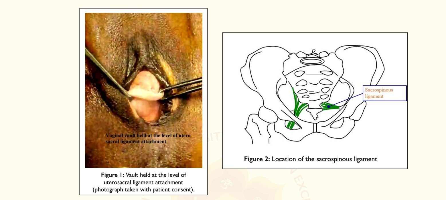
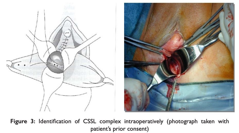
Patients were operated in a lithotomy position under spinal anesthesia. Vault was identified and marked with two Allis forceps, Fig 1. A longitudinal incision was given on the posterior vaginal wall to expose the rectovaginal space, on the right side of the patient, the epithelium was dissected laterally and the pararectal space was opened. After enterocele correction (if indicated), a window was created between the rectovaginal space and ischial spine by blunt finger dissection. Dissection was continued until the ischial spine was reached. Using the ischial spine as a landmark, the sacrospinous ligament was palpated, Fig 2. Then using 3 retractors peritoneum and the rectum were retracted and the sacrospinous ligament was identified, Fig 3. A delayed absorbable suture and non-absorbable suture was used for this procedure. The non-absorbable suture was placed with the help of a long 12 inches needle holder, through the sacrospinous ligament coccygeus muscle complex starting from superior border in an upside-down direction. It was taken 2 cm medial to the ischial spine, to avoid injury to the neurovascular bundle. A second stitch was taken 1 cm medial to the first stitch with delayed absorbable suture. Non-absorbable suture was passed through the vaginal vault full thickness except epithelium
and then tied by half hitch; the other end of the suture was then passed through entire thickness of vagina lateral to the first stitch, creating a pulley stitch. Absorbable suture’s ends were passed through vaginal wall full thickness on both sides.
Anterior colporrhaphy was performed, the upper vagina was closed, and lastly, the pulley stitch followed by reinforcing delayed absorbable stitch was tied. This was followed by perineorrhaphy, where required. Tying of sutures brought the sacrospinous ligament in direct contact with the vaginal epithelium and was pulled up.
When healing occurs, the vaginal epithelium is fused with the sacrospinous ligament and the vault remains suspended up. Postoperatively women were given broad-spectrum antibiotics for 5 days. Patients were followed up at 3 and 6 months, postoperatively. Data regarding quality of life, recurrence, complications, functional outcomes, urinary disturbances, bowel disturbances and sexual dysfunction were collected.
3. Results
During the study period of 7 months, a total of 8 patients were included. Mean age of the study group was 57.6 years, with a median parity of 2 children.
Table 1: Patient Characteristics
| Characteristics | Value | |
|---|---|---|
| Mean (SD) | Median (IQR) | |
| Demographic variables | ||
| Age (years) | 57.6 (11.7) | 56 (52.3-63.8) |
| Weight (kg) | 51.2 (4.9) | 50.3 (48.5-52.6) |
| Height (cm) | 151.4 (7.4) | 153.5 (149.3-156.5) |
| BMI (kg/m2) | 22.4 (2.1) | 22.9 (20.9-23.8) |
| Number of children | 2.4 (1.3) | 2 (2-3.25) |
| Reproductive history | ||
| Previous abdominal hysterectomy | 7 | |
| Previous vaginal hysterectomy | 2 | |
| POP-Q: Grade 4 prolapse | 5 | |
| Grade 3 prolapse | 3 | |
| Cystocele: Grade 2 | 2 | |
| Grade 3 | 2 | |
| Grade 4 | 3 | |
| Rectocele: Grade 1 | 5 | |
Among the 8 parous women, 7 delivered at home with help of untrained dais and one delivered in a hospital. Of the 8 studied cases, 5 had prior vaginal hysterectomy and 2 had prior abdominal hysterectomy. Additional surgeries performed on these patients included anterior repair in 6 patients and posterior repair in 5 patients.
None of the patients had peri-operative or early postoperative complications like pyrexia, urinary tract infections, or hematuria. During the follow-up period at 3 months and 6 months, the following symptoms were observed:
Table 2: Post-operative Outcomes
| Symptoms | Number of patients |
|---|---|
| Lower abdominal pain | 2 |
| Low back ache | 3 |
| Difficulty/ pain in urination | 1 |
| Dyspareunia | 1 |
No patients endorsed symptoms of pelvic pressure/bulge, difficulty or pain in bowel movements, urinary or bowel incontinence.
4. Discussion
The true incidence of vaginal vault prolapse after hysterectomy remains unknown. Long-term follow up of these patients would be required with not just a symptomatic questionnaire but also a clinical examination to look for compartment defects. In one 13-year study done on 2,670 patients the incidence of vault prolapse was found to be 0.4% but on pelvic examination, it was found to be 4.4% in all cases of hysterectomies. Further breakup of these data show that the incidence of vaginal vault prolapse is 11.6% when done for genital prolapse, as opposed to 1.8% when the hysterectomy was done for benign conditions [3].
Pre-operative prolapse of grade 2 or more is a risk factor for later developing vault prolapse. Urinary incontinence surgery, previous history of vaginal deliveries, and sexual activity are additional etiologic factors. Vaginal hysterectomy, however, is not a risk factor when done for conditions without pre-operative prolapse or factors related to pelvic floor weakness [15].
Postmenopausal women with surgically managed pelvic floor disorders are at increased risk of developing vaginal vault prolapse after a vaginal hysterectomy, as was seen in our case series. Six out of the 8 women had undergone a vaginal hysterectomy for genital prolapse and were also postmenopausal at the time of surgery. All the six women had a grade 3 or 4 prolapse. Five of them had home deliveries conducted by untrained traditional birth attendants and the parity of these women was greater than two.
As per a study to look at obstetric risk factors for genital prolapse, symptomatic prolapse increased by 3.3-fold in women who had 4 or more children as compared to those who had one child. Excessive stretching and tearing of the pelvic floor being the other predisposing factors while abdominal deliveries were protective [16].
Pelvic floor muscle dysfunction along with anal lacerations have been associated with pelvic organ prolapse. Body mass index, heavy occupational work, and low socioeconomic status are the other risk factors [8]. The women from our study were from a poor socioeconomic background, with poor nutrition, quicker return to heavy work after delivery, and poor spacing between pregnancies.
The incidence of women undergoing surgery for pelvic organ prolapse will rise, as epidemiological data suggests that women aged over 65 years will nearly double. Uterine prolapse is the main contributor to reproductive health problem that influences the women’s quality of life. The incidence of uterine prolapse is 7.6% in North India, approximately 20% in East India, and 3.4% in southern India i.e. Karnataka. The World Health Organization reports the prevalence of uterine prolapse to ranges from 2% to 20% in women younger than 45 years of age. 33% of the global disease burden in women is due to reproductive ill health [17].
Surgical techniques for advanced pelvic organ prolapse need to be individualized based on the defects in the various compartments. Suspension of the uterosacral ligaments at the time of hysterectomy has similar anatomic and subjective outcomes to sacrospinous ligament fixation at the end of 1 year and must be considered in all cases of advanced prolapse.
Though abdominal approaches may have increased long-term durability, when counselling their patients, surgeons should consider longer operating times and increased pain and the added cost of these procedures compared to vaginal surgery.
Vaginal vault prolapses occurring post-hysterectomy has several surgical modes of management as discussed above. There are no guidelines to date. Sacrocolpopexy is probably the most commonly performed surgery. The routes vary between abdominal and transvaginal, open, laparoscopic and robotic. Differences in repair may be with and without mesh, or with native tissue. Vaginal sacrocolpopexy was shown to carry a success rate of over 90% and was the most commonly performed surgery followed by abdominal sacrocolpopexy. It could also be combined with or without continence surgery depending on the symptoms. Each procedure has its own advantages and disadvantages.
In our case series, we performed unilateral vaginal sacrocolpopexy in seven women and colpocleisis in one. Sacrospinous ligament fixation (SSLF) entails the placement of sutures to the sacrospinous ligament, which is then attached to the vaginal vault. Its main advantages include shorter duration of surgery, shorter recovery time, and the avoidance of abdominal incisions
Vaginal sacrocolpopexy is a simpler approach. The technique provides maintenance of sexual function and achieves adequate vaginal length and width. It can also be combined with other reconstructive procedures and additional antiincontinence surgery [18]. It carries lesser anesthesia risk as it is done under regional anesthesia. The complication rate is low, with blood loss from injury to pudendal/inferior gluteal vessels, requiring transfusion in 4.3% patients. The other complications include nerve damage to the sciatic or pudendal nerves, dyspareunia, and injury to the rectum. Buttock pain is reported in 10–15% of patients, but usually resolves within 6 weeks. The fear of these injuries has resulted in more surgeons opting for abdominal route [19].
Abdominal sacrocolpopexy was associated with a longer operating time, longer time for recovery, and was more expensive than the vaginal approach. The trend towards a lower reoperation rate in the abdominal route compared to vaginal route is not statistically significant [18]. Laparoscopic sacrocolpopexy provides a good anatomical correction but the risk of general anesthesia; mesh erosion, and increased cost are significant disadvantages. By utilizing the vaginal pathway, this approach minimizes incisions and the likelihood of scarring [22].
The prevalence of pelvic floor dysfunction six years after primary surgery was quite high after primary POP surgery in a Swedish study. The incidence of urinary incontinence of 1 or more episodes per week was 41%, feeling of vaginal bulging 18%, and solid stool incontinence was 15%. Only 39% women were sexually active, of which 42% experienced dyspareunia, and 15% refrained from sexual activity due to discomfort or pain [20].
The primary aims of surgical treatment are the restoration of normal vaginal anatomy as well as improvement in vaginal bulge symptoms and the restoration/maintenance of normal bladder, bowel and sexual function. However, most of the studies use only the anatomical outcome as the primary outcome, with POP-Q stages I or 0 defined as the anatomical cure. A recent qualitative study based on patient interviews showed that women are most affected by the actual physical symptoms of prolapse (bulge, pain, and bowel problems) as well as by the impact that prolapse has on their sexual function. Failure of surgery is defined as the vaginal apex descending below the upper third of the vagina, or one of points Ba or Bp being greater than 0 cm, i.e. the anterior (Ba) or posterior (Bp) vaginal wall prolapsing beyond the hymen [21].
Our patients reported that their prolapse symptoms and quality of life were improved at 3 and 6 months after surgery. Unlike previous studies, their prolapse status was also measured by clinical examination before surgery and at 3 months after their operation. None showed a failure of surgery at the end of this study period. None of the patients had pelvic pressure symptoms or difficulty in bowel movements or incontinence post-surgery. Three patients had low backache, one patient had dyspareunia, one patient had dysuria, 2 of them had lower abdominal pain. Statistical significance of post operative complications could not be derived due to small sample size. We will continue to follow-up these patients for a period of 2 years to look at long-term outcomes.
5. Conclusion
Vaginal sacrocolpopexy is a safe and effective surgical technique for correction of PHVP. In the postoperative followup of 6 months for a few patients and 3 months for all patients, there was no persistence or recurrence of symptoms. Prophylactic steps during primary surgery can help to further reduce the incidence of vault prolapse.
Author Contributions: Conceptualization: Padmasri R., Keerthi A. V.; methodology: Padmasri R., Priyanka Holla P. N.; writing—original draft preparation: Priyanka Holla P. N.; writing—review and editing: Padmasri R.; visualization: Padmasri R.; supervision: Padmasri R
Funding: This research received no external funding.
Institutional Review Board Statement: Institutional Ethical Committee Clearance was obtained prior to data collection (Protocol Number: 04/2023).
Informed Consent Statement: Written informed consent was obtained from all subjects involved in this study.
Data Availability Statement: The data presented in this study are available on request from the corresponding author. The data are not publicly available due to privacy and ethical policy
Acknowledgments: Dr Kiran Ashok, Urogynaecologist, Dr Chandrashekhar YS, Urogynaecologist, Visiting consultants at Sri Madhusudan Sai Institute of Medical Sciences and Research
Conflicts of Interest: The authors declare no conflicts of interest
References
-
Post Hysterectomy Vaginal Vault Prolapse. Greentop Guidelines No. 46, RCOG/BSUG; July 2015.
-
Symmonds RE, Pratt JH. Vaginal prolapse following hysterectomy. Am J Obstet Gynecol. 1960;79:899–909.
-
Marchionni M, Bracco GL, Checcucci V, Carabaneanu A, Coccia EM, Mecacci F, et al. True incidence of vaginal vault prolapse. Thirteen years of experience. J Reprod Med. 1999;44:679–84.
-
Aigmueller T, Dungl A, Hinterholzer S, Geiss I, Riss P. An estimation of the frequency of surgery for post hysterectomy vault prolapse. Int Urogynecol J. 2010;21:299–302.
-
Brubaker L, Cundiff G, Fine P, Nygaard I, Richter H, Visco A, et al.; Pelvic Floor Disorders Network. A randomized trial of colpopexy and urinary reduction efforts (CARE): design and methods. Control Clin Trials. 2003;24:629–42.
-
Cruikshank SH, Kovac SR. Randomized comparison of three surgical methods used at the time of vaginal hysterectomy to prevent posterior enterocele. Am J Obstet Gynecol. 1999;180:859–65.
-
Margulies RU, Rogers MA, Morgan DM. Outcomes of transvaginal uterosacral ligament suspension: systematic review and meta-analysis. Am J Obstet Gynecol. 2010;202:124–34.
-
Braekken IH, Majida M, Ellström Engh M, Holme IM, Bø K. Pelvic floor function is independently associated with pelvic organ prolapse. BJOG. 2009 Dec;116(13):1706–14.
-
Gimbel H. Total or subtotal hysterectomy for benign uterine diseases? A meta-analysis. Acta Obstet Gynecol Scand. 2007;86:133–4.
-
Maher C, Feiner B, Baessler K, Christmann-Schmid C, Haya N, Brown J. Surgery for women with apical vaginal prolapse. Cochrane Database Syst Rev. 2016 Oct 1;10(10).
-
Freeman RM, Pantazis K, Thomson A, Frappell J, Bombieri L, Moran P, et al. A randomised controlled trial of abdominal versus laparoscopic sacrocolpopexy for the treatment of post-hysterectomy vaginal vault prolapse: LAS study. Int Urogynecol J. 2013;24:377–84.
-
Louis-Sylvestre C, Herry M. Robotic-assisted laparoscopic sacrocolpopexy for stage III pelvic organ prolapse. Int Urogynecol J. 2013;24:731–3.
-
Barber MD, Visco AG, Weidner AC, Amundsen CL, Bump RC. Bilateral uterosacral ligament vaginal vault suspension with site-specific endopelvic fascia defect repair for treatment of pelvic organ prolapse. Am J Obstet Gynecol. 2000;183:1402–10; discussion 1410–1.
-
Harmanli OH, Dandolu V, Chatwani AJ, Grody MT. Total colpocleisis for severe pelvic organ prolapse. J Reprod Med. 2003;48:703–6.
-
Dällenbach P, Kaelin-Gambirasio I, Dubuisson JB, Boulvain M. Risk factors for pelvic organ prolapse repair after hysterectomy. Obstet Gynecol. 2007 Sep;110(3):625–32.
-
Tegerstedt G, Miedel A, Maehle-Schmidt M, Nyrén O, Hammarström M. Obstetric risk factors for symptomatic prolapse: a population-based approach. Am J Obstet Gynecol. 2006 Jan; 194(1):75-81.
-
Harjit and Shehnaz Johnson. An Exploratory Study to Assess the Knowledge Regarding Uterine Prolapse and its Prevention among Staff Nurses in Christian Medical College & Hospital, Ludhiana, Punjab. International Journal of Development Research. 2019;9(01).
-
Toh VV, Bogne V, Bako A, Barrington JW. Management of recurrent vault prolapse. Int Urogynecol J. 2012 Jan;23(1):29–34.
-
Barrington JW, Edwards G. Post hysterectomy vault prolapse. Int Urogynecol J Pelvic Floor Dysfunct. 2000;11(4):241–5.
-
Crafoord K, Sydsjö A, Johansson T, Brynhildsen J, Kjølhede P. Factors associated with symptoms of pelvic floor dysfunction six years after primary operation of genital prolapse. Acta Obstet Gynecol Scand. 2008;87:910–5.
-
Nygaard I, Brubaker L, Zyczynski HM, Cundiff G, Richter H, Gantz M, et al. Long-term outcomes following abdominal sacrocolpopexy for pelvic organ prolapse. JAMA. 2013;309:2016–24.
-
Ciortea R, Roman MP, Mălutan AM, Bucuri CE, Ormindean CM, Nati ID, Mihu D. Comparison of laparoscopic sacrocolpopexy with vaginal reconstructive procedures and abdominal sacrocolpopexy for the surgical management of vaginal vault prolapse: a systematic review and meta-analysis. Frontiers in Medicine. 2023;10.
Disclaimer/Publisher’s Note: The statements, opinions and data contained in all publications are solely those of the individual author(s) and contributor(s) and not of SSSUHE and/or the editor(s). SSSUHE and/or the editor(s) disclaim responsibility for any injury to people or property resulting from any ideas, methods, instructions, or products referred to in the content.